����ժ Ҫ���������л��Ǽܣ�Metal- Organic Frameworks, MOFs����һ���ɽ��������л�������λ��װ���ɵľ������. �����л��Ǽܾ��п�϶�ȴ��ͳߴ�ɵ������������Ժá��ɷֿɵ�����������ε���Խ���ܣ��������������������Ҫ��Ӧ��DZ��. �������Ƚ����˽����л��Ǽ�����С����ҩ���������ҩ���ҩ��ĵ�����ϵ�Ĺ�������. ��������ܽ��˽�����MOFsҩ�������ϵ�������Ļ�ѧ���ơ����ѧ���ơ����������ơ��������ơ��������Ƶȷ����Ӧ�ý�չ. ������ܽ���MOFs���������Ʒ���Ľ�չ���ص�, ��չ����MOFs ����������������о���ս��Ӧ��ǰ��.
�����ؼ��ʣ��������л��Ǽ�; ҩ�����; ����; ���ѧ����; MOF-��������;
����Abstract����Cancer is a serious threat to human health and life safety. The number of cancer patients has been increasing worldwide. Therefore, it is of great significance to develop efficient cancer treatment methods. Currently, the main clinical methods for cancer therapy include surgery, radiotherapy, chemotherapy, immunotherapy, and so on. In recent decades, nanomaterials-based cancer treatment methods have shown great potential and value in radiotherapy, chemotherapy and immunotherapy. Metal-organic framework (MOFs) is a kind of material composed of metal nodes and organic ligands. The metal-organic frameworks have many advantages, such as large porosity, adjustable aperture and size, good biocompatibility, adjustable composition and surface modification, which make it promising in the field of cancer therapy. In this review, the methods for constructing MOFs-based drug delivery system (DDS) were first introduced. In particular, small molecule drugs can be efficiently loaded into metal-organic frameworks using non-covalent penetration, covalent cross-linking, and so on. At the same time, biomacromolecules-MOF system could be constructed based on the methods such as pore-penetrating and de novo approach. MOF-enzyme complex is more resistant to hush circumstances or protein inactivating agents than naked enzyme. Moreover, the metal-organic framework itself can be designed to be highly efficient nano-drug by using pharmaceutically active organic molecules or metal ions as ligands or metal ions. General surface modification methods were also reviewed due to their important roles for improving the solubility and stability of MOF-based DDS. In this reviewer, recent numerous researches on the cancer therapy of MOF-based DDS have been summarized. According to the type of treatment methods, the applications of MOF-based DDS in chemotherapy, photodynamic therapy, biomacromolecules-MOF based therapy, and other combined therapies (such as radiotherapy, and immunotherapy) for cancer were introduced, respectively. Finally, we summarized the advantages and challenges of MOFs in cancer therapy, and prospected the opportunities and development of this research field.
����Keyword����Metal-organic frameworks; drug delivery; chemotherapeutics; photodynamic therapy; MOF-proteins complex;
������֢����������������������в������Ľ�������˷�չ��Ч���������Ʒ��������ش���о�����[1,2,3]. Ŀǰ���ٴ�Ӧ�õ��������Ʒ�����Ҫ����������ѧ���ơ����������Ƶ�. ��Щ���Ʒ����ܹ���һ���̶��ϼ���һЩ������������ת�ƣ��Ӷ��ӳ����ߵ����������Ǵ����������ڳ��������������ԡ��������ܸ���������.
��������ʮ�������������ײ�������Ĺ⡢�硢��ѧ���ʼ����׳ߴ�ЧӦ���ص�[4,5,6]���������ײ��ϵ�����ҩ���Ƽ��ڼ������Ʒ���չ�ֳ��ɱ��������. ���������л��Ǽܣ�Metal-Organic Frameworks, MOFs����һ���ɽ��������л�������λ��װ���ɵľ�����ϣ����бȱ�������ͳߴ�ɵ������������Ժá��ɷֿɵ�����������ε��ŵ㣬���㷺Ӧ���ڴ���������������Դ�����ת����������Ϊ�����ҩ�����ϵͳ���ƽ̨����������������зdz����DZ��[7,8].
�����������Ƚ��������������л��Ǽ�����С����ҩ���������ҩ���ҩ��ĵ�����ϵ�Ĺ�������[9,10]��Ȼ���ܽ��˽�����MOFsҩ�������ϵ�������Ļ�ѧ���ơ����ѧ���ơ����������ơ��������ơ���������[11,12,13]�ȷ����Ӧ�ý�չ. ����ּ�ڹ��ɻ���MOFs��ҩ�����ϵͳ���������Ʒ�����ص�ͽ�չ������MOFs���о���ս��Ӧ��ǰ������չ��.
����1�� ����MOF��ҩ�������ϵ�Ĺ���
������ײ��������ܹ�����ҩ����Ӳ�������͵�ϸ���У�ͬʱ��ͨ����ɢ�����Ͻ����;��ʵ��ҩ����ӵ��ͷţ���˱��㷺Ӧ����ҩ������ϵͳ�������[14]. �����л��Ǽܾ��к��ʵĿ���������϶�ȡ��õ����������Ե������������ܹ�ʵ��ҩ����ӵĸ��غ͵���. ����ҩ������࣬�ò������ݴ�С����ҩ���������ҩ���MOFҩ������������н���.
����1.1�� С����ҩ��
�����ٴ��������������Ƶ�С����ҩ�������������ड������ʡ��װ����ʵ�. ����ͨ�����Ƴɿڷ�Һ����ע��Һ. ��ЩС����ҩ�����ױ������л������ҩ�������ʵͣ������Ҫ�����ҩ�����. ���⣬��Щҩ��û�����������ԣ��ᵼ�����صĶ�������. ���ڽ����л��Ǽܵ�С����ҩ�������ϵ������һ���̶��Ͻ����������.
�����ǹ��۽����ǽ����л��Ǽܸ���С����ҩ���һ�ֳ�����ʽ. �÷���Ҫ���ý����л��ǼܵĴ��϶�ͳߴ硢��ѧ�ȶ��Ժõ��ŵ㣬��MOF������ҩ�������Һ�У�ʹ��С����ҩ����������϶�У������ܼ�ȥ�����������С����ҩ��. ���ַ���Ҫ��С����ҩ��ijߴ���MOF�Ŀ��������������֮����н�ǿ������[15]. ���У���ҩ����Ӿ�������õ��ض�����λ����л������ſ������ҩ�������Ч��. ���磬Serre�����������ַ����ɹ��ؽ�����Ұ�����MIL��Materials of Institut Lavoisier��-100/ 101�ͼ1a����ÿ�����С�ĸ������ֱ�Ϊ92��56��ҩ����ӣ��Ӷ�ʵ�ָ�Ч��ҩ������[16]��Liu���˽��������र�����ZIF��Zeolitic Imidazolate Framework��[17]�����Ч�ʴﵽ51%.
����Ȼ�����ǹ��۽�����������Щ������л��Ǽ�û��ǿ����õ�ҩ�����. �Һã�������ӿ���ͨ�����۽������ֶν���ڽ����л��Ǽ��ϣ��Ӷ�������Ч��ҩ�������ϵ. ��һ����Ҫ������л��Ǽܺ�ҩ��������¼����ص㣺��1���������л��Ǽܱ����пɹ����۷�Ӧ�Ļ���λ�㣬�簱����ȩ�������������ŵȣ���2��ҩ��ͨ����������֮��������ҩ����ϵ�������ǰҩ�����ʣ���3������ҩ���ܹ����ض������������д��������л��Ǽ��ϱ��ͷų���. Wei����ͨ���γ�ϯ���ķ�ʽ�����а��������ŵ�DOXҩ�����������л��Ǽ��ϵ�ȩ�������Ź��۽������Ӷ�ʵ��ҩ������ڽ����л��Ǽ��ϵĸ�Ч���أ�ͼ1b������������pH�������ͷ�ҩ��[18].
����ͼ1 �������ڽ����л��Ǽܵ�С����ҩ������ϵͳ
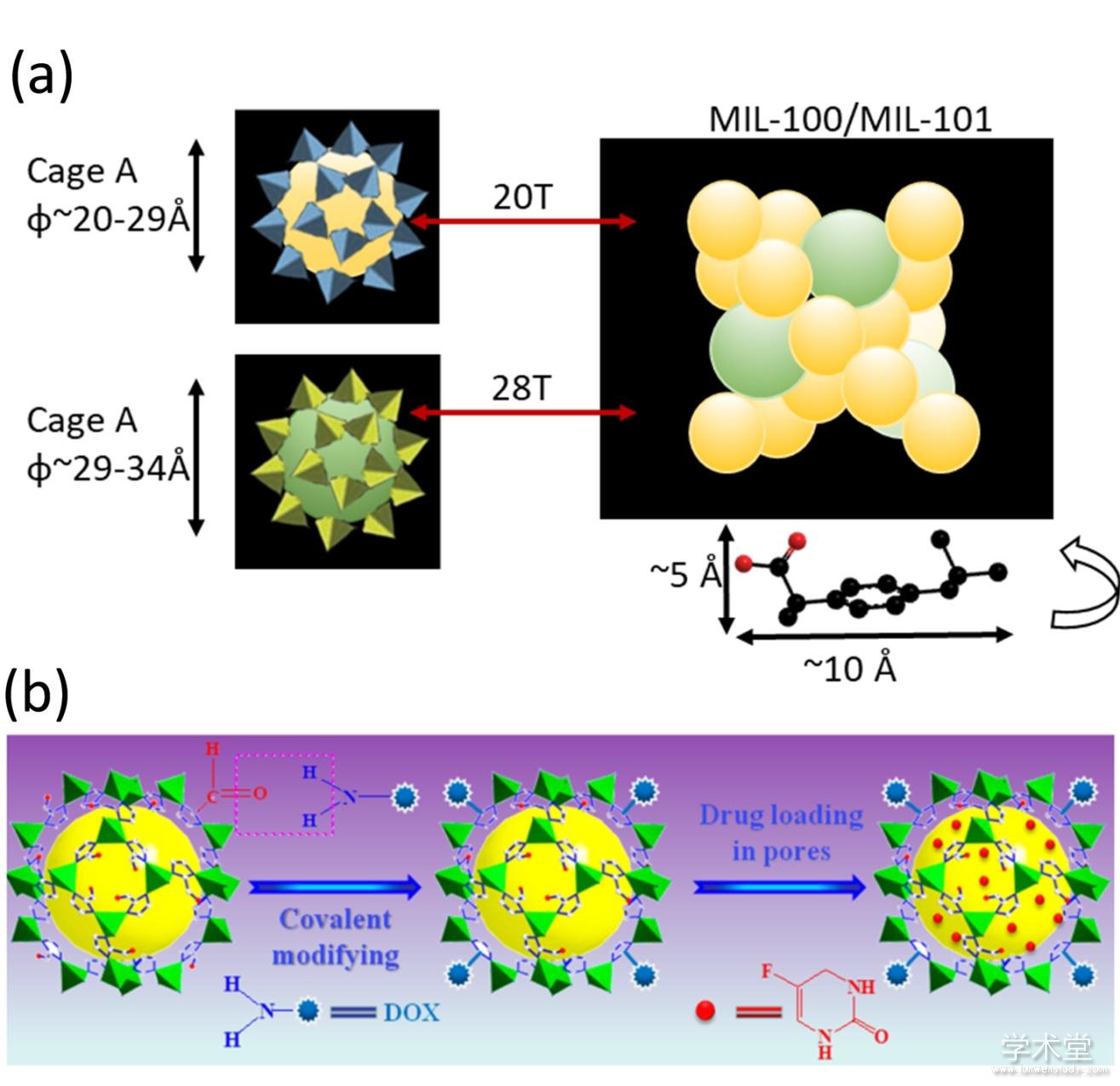
����Fig.1 Illustration for construction of MOF based small drug molecular delivery system
����(a) MIL-100��MIL-101�Ľṹʾ��ͼ[16]��(b)ҩ����ZIF-90���۽�������[18]
����1.2�� ��������ҩ��
������������ҩ����Ҫ�������ġ������ʡ����塢����������. ��Щ�������ӱ������ѽ���ϸ���������Ҫ�ض������彫���������ϸ�����ӹ���[19]. �����л��Ǽܾ������õĿ�ܽṹ��ƿɱ��Ժ��ɱ���ı���ɵ��ԣ���һ�ַdz���DZ������������ҩ�����ƽ̨�������ǵ�����ҩ��[20,21]��. Ŀǰ������MOF-��������ҩ���Ѿ������౨��[22,23]������MOF-�����ʸ�������о���Ϊ�㷺.
����MOF-�����ʸ�������Ҫͨ��������������л��Ǽ�֮���γɼ���Ǽ���ʽ�������. MOF-�����ʸ�������γɷ�ʽ���Է�Ϊ������̶������۽�����������ԭλ�������ȷ�ʽ.�ڹ��۽������У����õ����ʵİ����Ȼ���������л��Ǽ��ϵ���Ӧ�����ţ����Ȼ������۽������ܹ�ȷ���ϸߵĵ����ʸ���Ч�ʣ����ұ��⵰��й¶[22]. ���˹��۽������⣬������ʽ��Ҫ���������л��Ǽܺ͵�����֮��ķ��»�����������������. ���У�����̶�����ʵ�������dz��ºͣ������ڱ��ֵ����ʵĽṹ�����[24,25,26]. ��������������ԭλ�������ķ�ʽ�dz��ܻ�ӭ����ϸ�������£�
����1.2.1�� ����
����MOF-�����ʸ������������ÿ����ķ����ϳ�[27,28,29,30,31,32,33,34]. �������ӵĿ�������������������ʿ��Ľ����л��Ǽܻ�Ϸ�����ʹ�õ����ӽ�������л��ǼܵĿ�϶��. 2011�꣬Ma�����״ν��ߴ�Ϊ3.3 nm× 1.7 nm× 1.1 nm����������øMP-11ͨ��������������������л��Ǽ�Tb-mesoMOF�У�ͼ2a��. MP-11������Ҫ�������Ŀ��У���0.9 nm�Ŀ����ÿ���ΪMP-11�ĵ�������ṩͨ����������������ø���. ʵ������������MP-11���Է�ֹMP-11�ۼ�ʧ��[35]. ����Դ��ڽ����л��Ǽܿ�����ķ��ӣ����磺ϸ��ɫ��C��Cyt c����?2.6 nm × 3.2 nm × 3.3 nm����Ҳ���Խ��뵽�����л��Ǽ���. �����о���ʾϸ��ɫ��CҪ����һ����Ĺ��ͱ任�ſ��Խ��뵽��Խ�С�Ľ����л��Ǽܿ�϶�У�ͼ2b��[36].
��������ڽ����л��ǼܵĿ��ɵ����Լ�һϵ�к�������ƣ�Zhou���˿������������ֲ�ͬ�ߴ��϶�Ľ��MOF��PCN-333��[37]��ʵ�ֵ����Ӱ������ֲ�ͬ�ߴ�ĵ���ø. ����ø��ʹ�û���ͨ���Ƕ��ģ���ˣ��о��߿�����PCN-333ø�������ڲ�ͬ�����еĴ����ԣ��������PCN-333ø�������ڸ��ֽ����еĻ��Ծ�����¶øҪ��. ������PCN-333�Ŀ��в�������Ծۼ������»��pHʧ������. PCN-333����ø�����ܹ�����ø�������ã�������ʵ��ø�Ŀ�ѭ��ʹ�ã�����һ����Ȳ��ϴﲻ����Ч��.���Ƶģ�Zhou���˽��������ֿ�϶�Ľ����л��Ǽ�PCN-888����װ�����ֲ�ͬ�ߴ��ø[38]. PCN-888���Ŀ���װ��һ��������������ø��GOx�����ӣ��еȴ�С�Ŀ���װ��һ������������ø��HRP�����ӣ���С���ǿյģ�����Ϊ���������ͨ����ͼ2c��. ����øֻ�ܰ���˳����뵽PCN-888�Ŀ�϶�У��Ȱ���GOx�������HRP���ӣ�����һ��ø�Ĵ�������Ӧ��. ����ϵ�������кܸߵ�ø�����ԣ������Ա���ø������ø����.
�������˽�ø�����ڽ����л��ǼܵĿ����⣬��Һ����[39]��������������ø�����ڽ����л��ǼܵĿ���. ��Һ�������ǽ��ϳɺõĽ����л��Ǽ�ͨ��һ����Һ��ʹMOF��ø�ı�������һ����ά�Ľ����. �÷����ɹ�ʵ���˴������ø�İ��������Թ�������øΪģ�����Ĺ�������ø-MOF��������иߵ�ø�����ԡ��ȶ��ԺͿ�ѭ������.
����ͼ2 ���������������ʷ���
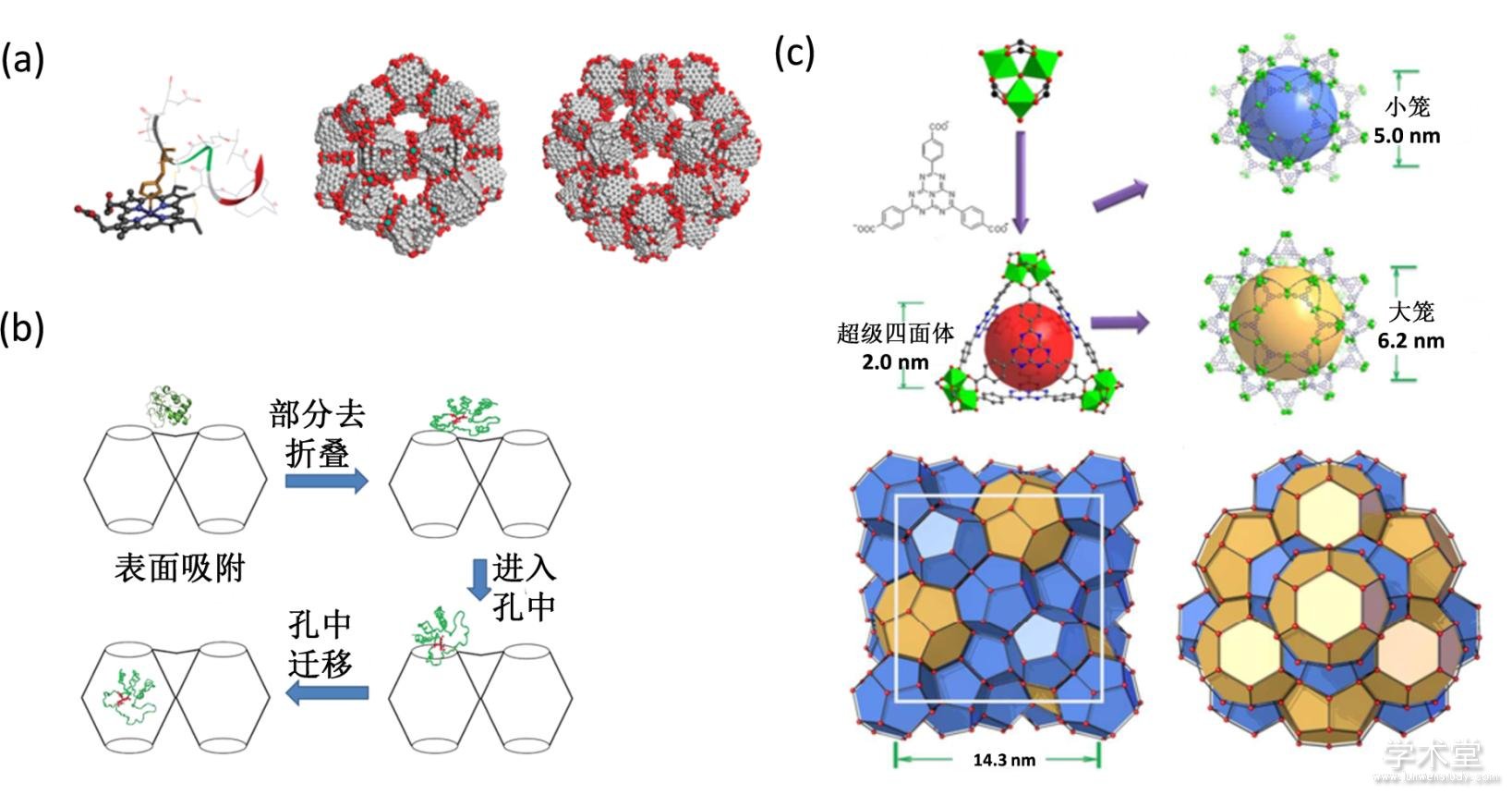
����Fig. 2 Proteins penetration into MOF
����(a) MP-11�ķ��ӽṹ��Tb-mesoMOF�Ŀṹ[35]��(b) Cyt c����Tb-mesoMOF��Ѩ�Ļ���[36]��(c) PCN-888�Ľṹʾ��ͼ[38]
����1.2.2�� ԭλ������
���������л��Ǽܵ�ԭλ���������ֳƹ�����������һ��ֱ���ڵ����ʵ�������ӱ������������л��Ǽܵİ�������. �����Ŀ�������Ҫ������л��ǼܵĿ׳ߴ��뵰���ʵijߴ��ƥ��. ���֮�£�ԭλ�����ķ���������������Dz����Ƶ����ʷ��ӵijߴ�[40,41,42].
��������øͨ�����л�����֮�����������ã����������ʹ�����ߵĽ�ϳ�Ϊ����. �������л��Ǽ�ZIF-8��ԭ���뵰��ø��������Һ�л�ϣ�һ��ʱ��ɻ��ZIF-8ø���������Ч��ʵ�ֵ���ø��ԭλ������ͼ3a��[43]. ͨ�����ַ����õ���ZIF-8ø������ͬ������ά��ø��������£�����ˮ�������ˮ���л��ܼ��ȣ��Ļ���. ͬʱ������ZIF-8��ƫ���Ի����лᱻ���⣬���ZIF-8ø���������ʵ�ֵ�pH�µ���ø�Ŀɿ��ͷţ��Ӷ��������Ӵ������Ӵ�����. ����MOF����ZIF-90��Ҳ����ͨ������ԭλ�����ķ�����������ø����[44].
������ø����ԭλ����MOF�ķ�����Ȼ���ܳߴ�����ƣ������ܵ���ø�ı������Լ����滯ѧ���ʵ�Ӱ��. �Ե��ױ���İ�������л�ѧ���ο�����Ч�Ŀ���ZIF-8�ڵ��ױ��������[45]. ���ױ����������л�������������Ӧ�����ӱ��渺��ɣ��ٽ�ZIF-8�������������ױ����Ȼ��������Ҷ�����Ӧ�����ӱ�������ɣ��谭ZIF-8������ͼ3b��. ����ģ��Ľ���������ı����ɴ��Ӱ��������������������. �ɼ����ı����ﻯ���ʶ���ʵ��ԭλ�����dz���Ҫ. �������Ƶ�ԭ�����ڵ����ʱ������ΰ�������ʹ�������кܶ�İ��װ���л������ٽ�ZIF-8�������������ͼ3c����������������������Ľ����л��Ǽܣ���HKUST-1���ڵ����ʱ��������[46].
�������˵������ʶ�ԭλ����Ч����Ӱ��֮�⣬�����л��Ǽܿ����ʶ��������еĵ���ø�Ĺ���Ҳ����Ӱ��. �о���������ø��������ˮ�ԵĽ����л��Ǽ�MAF-7����ZIF-90�п����ڸ��¡�ʧ������л��ܼ��Ȼ����б��ֻ��ԣ����ǰ�������ˮ�Խ����л��Ǽ��еĹ�������ø����ø���ʧ�ͼ3d��[47].
����ͼ3 MOFԭλ���������������ʷ���
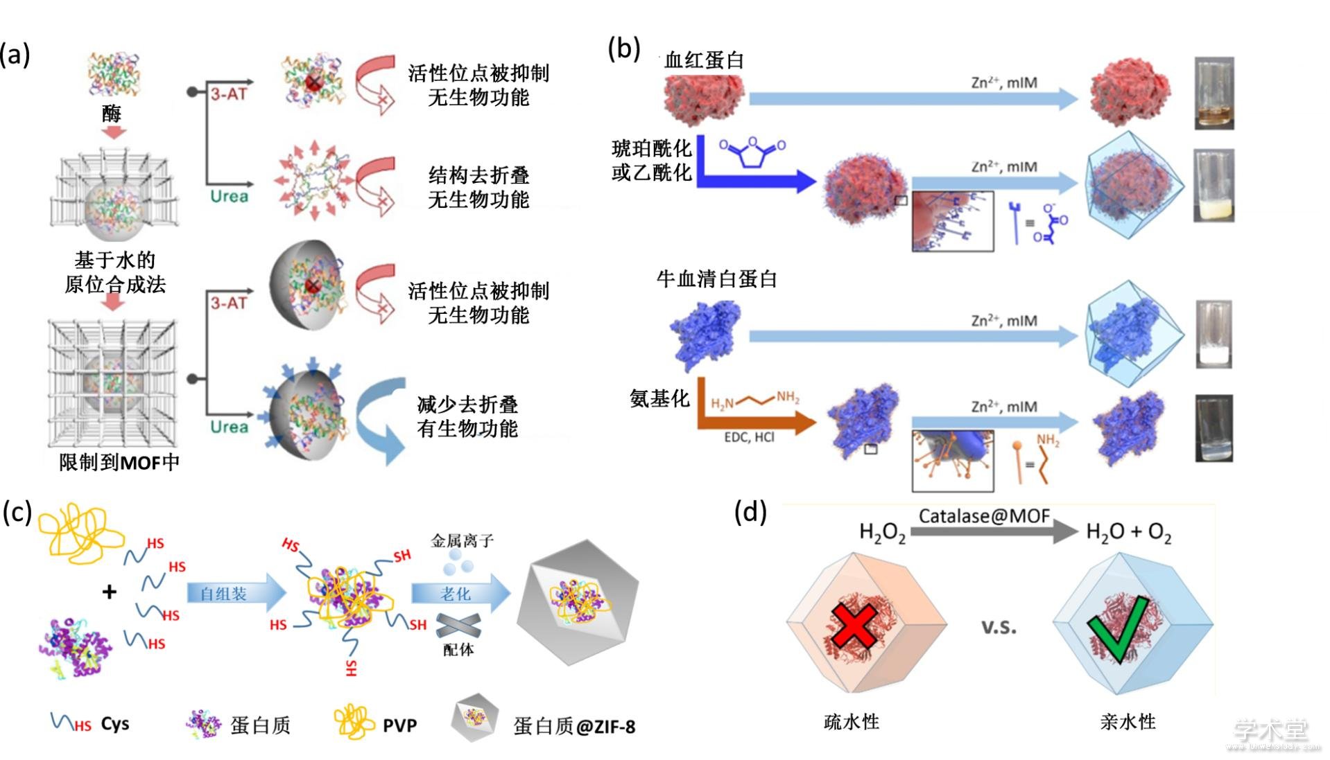
����Fig. 3 De novo approach for construction of MOF-protein complex
����(a) �������ϳɷ���������øʾ��ͼ[43]��(b) ���ױ������ζԹ�����������Ӱ��ʾ��ͼ[45]��(c) ������ٽ���������ʾ��ͼ[46]��(d) ��ˮ�Ի�����ˮ�Խ����л��Ǽܰ���øʾ��ͼ[47]
����1.3�� �����л��Ǽ�������Ϊҩ��
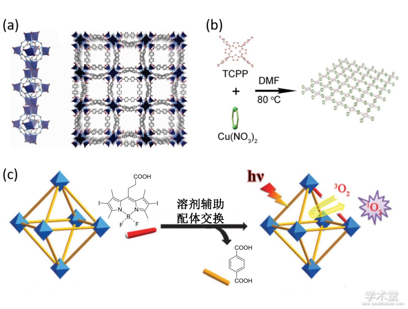
����������ϵ���ǽ�С����ҩ�����������ҩ�����ڽ����л��Ǽ������֮�⣬��Щ�����л��Ǽ�����Ҳ������Ϊҩ��. ���ַ���ֱ��ѡ�þ���ҩ����Ե�������Ϊ�����������ӣ�ֱ����װ�ɽ����л��Ǽܲ���ʵ�����ƹ���. �÷������������ҩ����ӵ�ˮ���ԣ����ҿ���һ���̶��Ͻ���ҩ������ﶾ��[48].������ҩ�﹦�ܵķ�����Ϊ�����γɿɽ����л����ҩ����ϵ.���磬Rosi���˽���Դ�Ե���������Ϊ���壬�����������װ�ɽ����л��Ǽܣ����ָ÷����ܹ�ά������ԭ�е�������ԣ�ͼ4a��[49,50,51]. ���⣬Wu�������þ��й����Եķ�����Ϊ����ϳɽ����л��Ǽܣ�ͼ4b��. �ڸ���ϵ�У��������ڽṹ�����б��������п����������˹��������Ӿۼ��������⣬�������������ˮ�Թ�����������ˮ��Һ�е��ܽ���[52]. ���ڹ���������Ľ����л��Ǽ�ͬ�����й����ԣ��ڹ��������¿��Դ���������������. ͬʱ�����ý����л��Ǽܵijߴ�ɵ����ʣ�ͨ�����ƺϳ��������Ի������ߴ�Ľ����л��Ǽܣ����Խ�һ�����������Ч��[53]. ������ֱ����Ϊ���幹�������л��ǼܵĹ�������Xie���˷�չ��һ���µķ�����ͨ���ϳɺ����彻���ķ�ʽ�ϳɹ����Խ����л��Ǽ�[54]��ͼ4c��.
����ѡ�þ���������ԵĽ����ǹ��������л����ҩ����ϵ����һ��;��. ����п�����ȱ��㷺���ڿ�������������Ϊ���������װ�ɵĽ����л��Ǽ��ڿɿص������·ֽ��ͷų��������ӿ���ʵ�ֿ���Ч��[55,56,57,58]. һЩ�ܹ����ϸ���Է��������жȵĽ������ӣ�����������ٵȣ�����ʵ�������װ�ɽ����л��Ǽܿ������ڷ�����������������߷���������Ч��[59]. ���⣬�����š��̵���Ϊ���������װ�ɵĽ����л��Ǽ�Ҳ����������ҽѧ����[60,61,62,63].
����ͼ4 �ϳ�MOF����ҩ��
����Fig. 4 Construction of MOF-based nanodrugs
����(a) п-�����ʽ����л��Ǽ�ʾ��ͼ[50]��(b) ͭ-TCPP�����л��Ǽ�ʾ��ͼ[52]��(c) UiO-PDT �������ĺϳɺ�Ӧ��ʾ��ͼ[54]
����1.4�� �����л��Ǽܵı�������
������������һЩ���ԣ���ߴ硢��ɡ�������ˮ���Լ�������������ʺ��ܶȵȣ���Ӱ����������ѪҺѭ����˥�ڡ�����ֲ��Ͱ�������. ���У��������ij�ѪҺѭ������ͨ���dzɹ�ʵ�ְ���ҩ����͵�һ����Ҫǰ��.
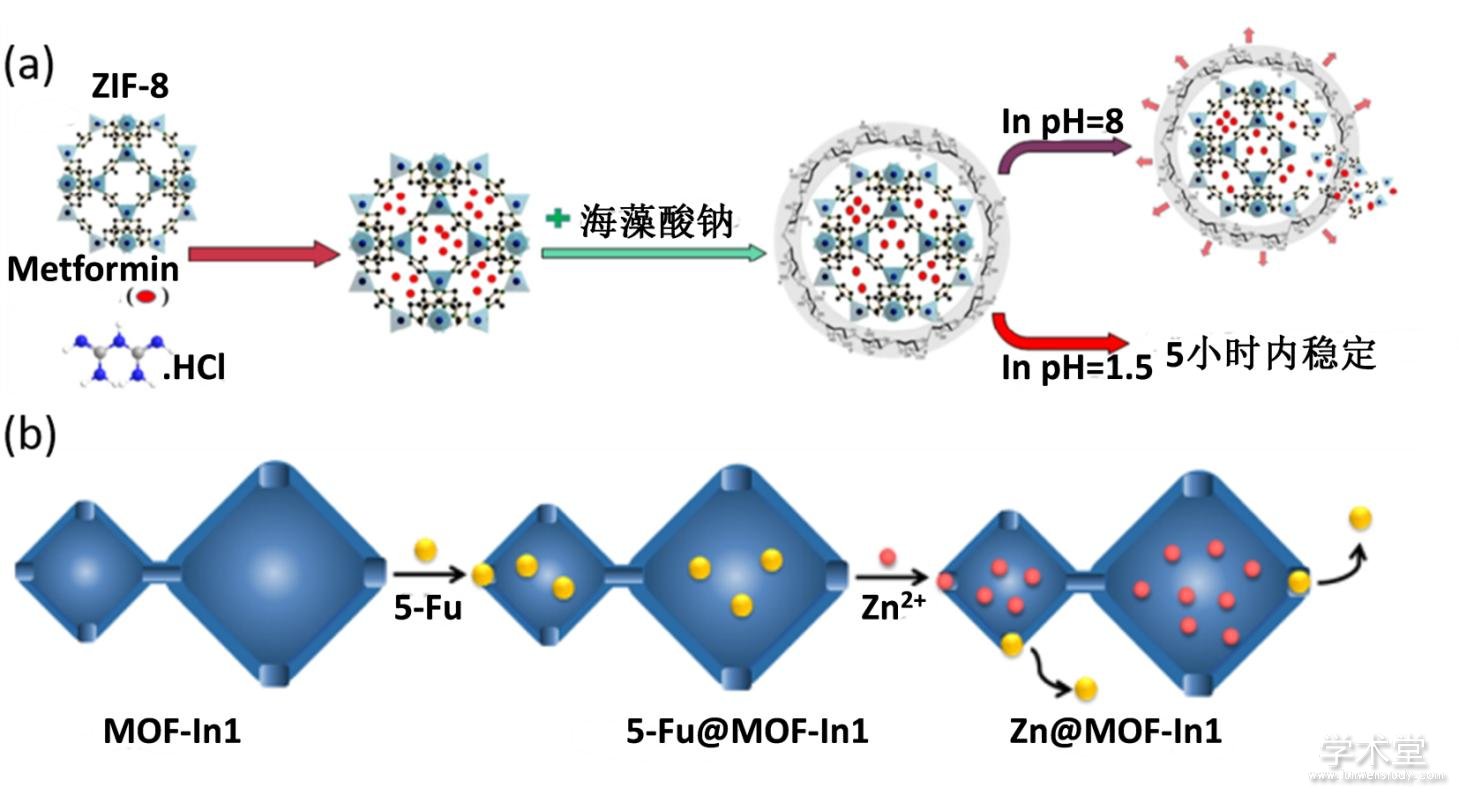
����Ϊ��ʵ�ֽ����л��Ǽܲ��ϵij�ѪҺѭ����Forgan���˽���ˮ�Եľۺ���[64,65,66]������Ҷ�����������Ϊ�������ı���������ϣ��Ӷ��������������ˮ�Բ�����ѪҺ�е��ĵ��������ã�����������������ϸ�����ɣ�ͼ5a��. ����֮�⣬�ڱ������PVP���������ײ������γ����ֶΣ�ͬ�������ڽ����л��Ǽ���ø��������ϵ[67]. ���⣬��MOF-��������������ϸ��Ĥ�������Ա������ײ������⣬�����Ա���ϵͳ�����������ͼ5b��. ����ϸ��Ĥ������������ʣ�����ϵ������ʵ��������λ��ͬԴ����������ϸ�����ͷŵ���ҩ��Ӷ�����������������������������Ч��[68,69]. ���⣬��MOF������������ϣ���CaCO3��,Ҳ������߲��ϵ��ȶ��Բ�ʵ��ҩ��Ŀɿ��ͷ�[70].
����ͼ5 MOF�ı�������

����Fig. 5 Surface modification of MOFs
����(a) �����л��Ǽܱ�������PEGʾ��ͼ��pH����ҩ���ͷ�[66]��(b) �����л��Ǽܱ������ϸ��Ĥʾ��ͼ[68]
����2�� ����MOF��ҩ�������ϵ��Ӧ��
���������л��Ǽ�����������������о��DZ��������ȡ��һϵ�н�չ. �������Ʒ�ʽ�����𣬸ò��ֽ�չ��Ҫ�ɷ�Ϊ�����ĸ����棺
����2.1�� ��ѧ����
���������л��Ǽ���Ϊ��DZ���Ļ�ѧ����ҩ��������壬���м��¼�����������1���״�С�ɵ��Ժͻ�ѧ���ʿɵ��ԣ���2��������ֵ�����࣬����ʵ�ֱ��������Σ���3�����BET�ȱ���������ڴ�������������ӣ���4��һ�ֽ����л��Ǽܿ��м��ֲ�ͬ�ߴ�Ŀף���ʵ��ͬʱ���ؼ��ֲ�ͬ���ܵ��������. ҽѧ�����Ӧ�öԽ����л��Ǽ���ҩϵͳ�����и��ߵ�Ҫ��. Ϊ��ȷ����Ч��ҩ����ͣ�����Ҫ�������л��Ǽܾ���һ�¼������ԣ���1����Ч�ĸ���ҩ�������������Է��ӣ���2�����ڱ������λ���أ��Ӷ�ʵ�ְ����Եص���ҩ���3���ɿص�ҩ���ͷţ���4�����ϱ����ɱ������л�����ⶾ������.
����2006�꣬Serre�����״ν������л��Ǽ�MIL��������ҩ������[16]. ����ϵʹ�õ�MIL-100��MIL-101���ɽ�������Cr��������������Ͷ��������������װ���ɣ����нϴ�Ŀ���25—34 ?�����ȱ������3100--5900 m2g-1���Ϳ������2 cm3g-1��. �����ڸ߱ȱ�����Լ���ҩ�����֮�������������ã�MIL-101��ҩ��װ����Զ����һ��ķ�ʯ���ײ���. �ݱ�����ÿ��MIL-101����װ��ҩ��ߴ�1.376 g����ÿ��MIL-100����װ��0.35�˲����ҩ��. ���⣬����MIL-100��MIL-101�Ļ�ѧ�ȶ��Էdz��ߣ����ֲ��Ͼ����нϳ��Ŀɿصĸ�ҩʱ�䣬Ϊ�����л��Ǽ�����ҩϵͳ�е�Ӧ���ṩ�˼��������.
��������һ������л��Ǽ�ZIFҲ��֤�����к�ǿ��С����ҩ���������. ���У�ZIF-8��һ���н���п��2-��������װ���ɵĽ����л��Ǽܣ������㹻��Ŀ�϶�Ϳɿص���ò. 2016�꣬Liu�鱨���˵�һ������ҩ�︺�����Ͱ�������������һ���Ļ���ZIF-8�Ŀ�����ҩ���������व�������ϵ�����ɹ�ʵ��������������[17].
���������������������pH�����̼�ҩ���ͷŶ���ʵ��ҩ��Ŀɿ��ͷž�����Ҫ����. Panahi��������ZIF-8��������˫��ҩ����ӣ�ʹ��������ϸ���ڵ�pH�̼����ͷ�ҩ�����[71]��ͼ6a����ʵ����ϸ����Դ�����Ӽ����ҩ���ͷ�. ����ZIF-8֮�⣬Yu���˻����������������л��Ǽ�����������������ҩ����ӣ���ʵ����pH�ɿ�ҩ���ͷ�[72,73]. ���磺��4,4'-�������������ᣨH2SDBA����N,N-������������DMA�����������װ���ɵĽ����л��Ǽܿ���ͨ���Ľ������������ष���[74]���ڵ�pH�������ͷ�ҩ����ӣ��ܹ����������˹�����ϸ��ϵ������. �������ϵ�У�pH������ҩ��������ȫ�ͷŵ�ʱ���ԼΪ20Сʱ. �����ʾ�������ष���λ�ڽ����л��Ǽܵ�ͨ���м䣬ͨ���������2.991 ?������-������2.915 ?��֮�������ý��. ��������������֮�⣬��������������ı���֮����π-π����ã�����Ϊ3.370 ?. �⼸���������ʹ���������ऺͽ����л��Ǽ�֮��ǿ�����Ľ���������Ӷ�ʵ��ҩ��Ļ���.
������������һЩ���ڽ����л��ǼܵĿɿ��ͷŵ���ҩ��ϵ������������������ϸ��������.���磺Wang���˷�չ��һ�ֻ���п���ӵ�pH�ɿ��ͷŵĽ����л��Ǽ�[Zn3(BTC)2(Me)(H2O)2](MeOH)13 (H3BTC = 1,3,5-benzenetricarboxylic acid, Me = melamine)[75]. ��Щ�����л��Ǽܰ���ҩ��ķ�ʽ����ͨ���Ľ���ʵ�֣�ʹ��ҩ�����ͨ�����϶������ö��������ڿ���. �����ķ�ʯ������л��Ǽ�ZIF-90����п���Ӻ�����2-ȩ��������װ���ɣ����д�����ȩ��������ͨ��ϯ��Ӧ���������DOXҩ����ӹ��۽���. ͬʱ��ZIF-90���ܹ�����ҩ������������ण��Ӷ�ʵ�����ֿ�����ҩ��Ĺ�ͬ����. ��pH 5. 5�Ļ����У�ҩ����16Сʱ֮���ͷ����ﵽ95%���������Ի�����ҩ���ͷ������Ž���[18].
���������л��Ǽ�ҩ�������ϵ���˶�pH�̼���Ӧ֮�⣬�����������أ���п���ӣ���Ӧ�ͷ�ҩ��. Yang���˽���������װ�ص������л��Ǽ�MOF-In1�У�ͼ6b��. ����MOF-In1�Ŀ�϶�Ǹ���ɵģ���ϸ���ڵ�п����Ũ�Ƚϸ�ʱ��п���ӻ���뵽��϶��. �ھ��������£���������ҩ�ﱻ�ͷų���[76].
���������л��Ǽܻ��ܹ����ڵ�������ҩ���DOX[77,78,79]��ϲ����[80]����ɳ̹��valsartan��[81]��������[82]�ȣ����ﵽ�˱�С����ҩ����õ�����Ч��.
����ͼ6 �����л��ǼܵĴ̼���Ӧҩ���ͷ�ʾ��ͼ
����Fig. 6 Stimulus triggered drug release of MOF
����(a) �����л��Ǽ�pH��Ӧ��ҩ���ͷ�ʾ��ͼ[71]��(b) �����л��Ǽܵ�п������Ӧҩ���ͷ�ʾ��ͼ[76]
����2.2�� ���ѧ����
�������ѧ���ƣ�photodynamic therapy, PDT���ǽ����귢չ������һ���������������ֶ�[83,84,85,86]����Ҫ�����Ͷ��ԵĹ������ڹ��������´���������������ɱ������ϸ��[87,88]. ��ͳ��С���ӹ���������ˮ���Բ���ȵ͵�ȱ�㣬�������ٴ�Ӧ������. ���������ײ���������������ˮ���ԡ����︻�����ܵ��ŵ㣬�����ֲ���ͳС���ӹ������IJ��㣬������������ѧ���Ʒ�����о��DZ��[89,90]. ���У������Խ����л���ܣ���������ΪС���ӹ����������廹�DZ������й����ԣ��������ܽṹ�����ɵ��ض����㷺���������Ĺ��ѧ�����о�[91,92,93,94]. �����л��Ǽ�Ӧ����PDT����Ҫ�ŵ����ڣ���1��������ɿصľ���ṹʹ�ñ�����������еĹ��������ӱ˴˷ֿ��������˹������ۼ���𣬺ܴ�̶��������ڻ������IJ�������2���ṹ�����ڵ���̬������ɢ����3�����׳ߴ�ЧӦ��������˹�������ˮ���ԣ���ǿ�˱�ϸ����ȡ��������4�������л��Ǽܿ����オ�⣬�������õ�����������.
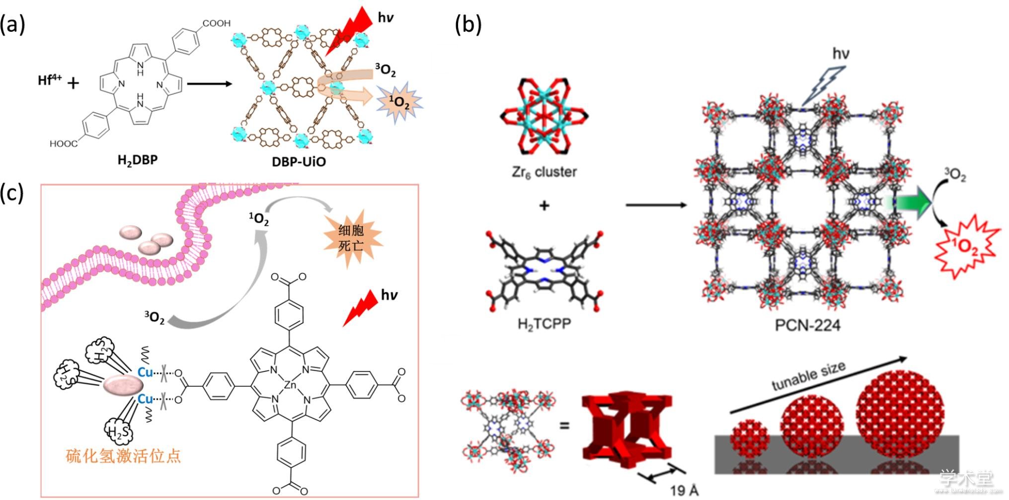
����߲�����������������صķ��ӽṹ���ɱ�������Ϊ���������������װ�ɽ����л��Ǽ�. ������һ�ص㣬Lin����2014�걨���˵�һ��������PDT������߲�������л��Ǽ�[95]�����ɹ�����Ӧ���������Ļ������ƣ�ͼ7a��. �ڸ���ϵ�У�߲��������5,15-di(p-benzoato)porphyrin (H2DBP)���������λ��װ�ɽ����л��Ǽ�DBP-UiO���ҹ������ĺ����ߴ�77 wt%����������̬��������H2DBP���������ϣ��������ڱȽ��������Ƶİ�֢. ����ģ�����ƽ��˵��DBP?UiO�ڹ�����²������ԣ��ﵽ���������������������������Ч��. ֵ��һ����ǣ��ڹ��ѧ����֮��û�г���С��Ƥ������֯����. ��һ������֯����ѧ����ע��DBP?UiO��ʵ����������λ�о���ϸ�����룬˵������������ϸ����������. ��DBP?UiO֮���о�С�鱨���˻���߲�ԵĽ����л��Ǽ�DBC-UiO��ʵ���˱�DBP?UiO���ߵĵ���̬������Ч�ʣ������ɹ�Ӧ���ڽ᳦���Ļ�������[96].
�������˽����л��Ǽܵijɷ�֮�⣬�����л��Ǽܵijߴ�Ҳ��Ӱ������ѧ����Ч����һ����Ҫ����. ͨ��ɸѡ����ߴ�Ĺ����Խ����л��Ǽ���������������������İ����������Ӷ������PDTЧ��. Zhou�����������߲����װ�Ľ����л��Ǽ���֤����һ���루ͼ7b��. ���Ǻϳ��˼��ֲ�ͬ�ߴ磨�ֱ�Ϊ30��60��90��140��190 nm���Ĺ����Խ����л��Ǽܣ���ϵͳ����ߴ��PDT����Ч����Ӱ��. ����ϸ�������о������90 nm�Ľ����л��Ǽܵ�����Ч����ѣ�˵��90 nm�Ŀ�����������ijߴ�[53].
��������PDT����Ч����ͨ���ⲿ�̼��ͻ���ʵ��PDTҩ��Ŀ���Ҳ���зdz���Ҫ������.����̬���IJ����벻������䣬���ͨ�������л��Ǽܵ�����ɿ���ʵ�ֹ���п���ʵ��PDT���Ƶĵ���. Zhou���˽���ת�����ط���1,2-bis(5-(4-carbonxyphenyl)-2-methylthien-3-yl)cyclopent-1-ene (BCDTE)�����п��װ��˫ɫ�����Խ����л��Ǽ�[97]. ����������£���ת������BCDTE�ػ���������TCPP����ĹⱻBCDTE���գ���������̬���������ڿɼ������£���ת�����ط���BCDTE�������������������ã���������̬��. ����ϵʵ������ϸ���ڿ�����Ƶ���̬���������Ӷ�ʵ����PDT����Ч���Ŀ������.
��������������־������⣩��������Խ����л��Ǽܵ�PDT����Ч������һ�ַdz���Ч�IJ���. ���磬Tang���˽�ͭ������п߲����������װ�ɽ����л��Ǽ�NP-1��ͼ7c����ͭ�����ܹ���ȫ���п߲�������ӫ�⣬�Ӷ����ؽ�������ĵ���̬������[98]. �������л��Ǽ��������ϣ�ͭ���ӽ����ڵ�������ⷴӦ������ͭ��ʹ�ý����л��Ǽֽܷ⣬�����ӫ����֮�ָ�������̬���IJ�������������Ӷ�ʵ�����⼤��Ĺ��ѧ����. ����ϵ�����ڽ�ֱ�����Ļ�����ѧ���ƣ����ƺ������ӽ���ȫ����.
������߹��ѧ���ƻ�����ͨ������ϸ���ڵĻ�ԭ��������ʵ��. ���磬��ԭ�Թ����ģ�GSH�������Ĺ������ڹ����´������ĵ���̬����һЩ�о�������������Щ�����ķ��ӿ���ȷ�������ĵ���̬�����������ģ��Ӷ������߹��ѧ���Ƶ�Ч��. ���������л��Ǽ� {CuL-[AlOH]2}n (MOF-2, H6L=mesotetrakis(4-carboxylphenyl)porphyrin))�ܹ�����������̬�������������ķ���[99]. �ý����л��Ǽܱ����̽���ϸ�����ڹ��������²�������̬��������̬����ͨ����϶��ɢ��ϸ����. ϸ���д�����GSH�ᱻ�����л��Ǽ��������������ɢ��ϸ���еĴ����ĵ���̬����ȷ���˵���̬���Ļ��ԣ��Ӷ���߹��ѧ����Ч��. ����ϵ����Ч��������ҵ�������ٰ�ҩ��ϲ��������.
����ͼ7 MOF���ڹ��ѧ����
����Fig. 7 MOF-based photodynamic therapy
����(a) Hf-DBP�ϳ�ʾ��ͼ�͵���̬����������[95]��(b) PCN-224�Ľṹ�͵���̬�����������Լ��ߴ�ɵ�ʾ��ͼ[53]��(c) MOF NP-1�͵���̬������ʾ��ͼ[98]
����2.3�� ����MOF-����������ϵ�����Ʒ�ʽ
����MOF-�����ʸ�����������ҽѧ����͵����ʹ����������ŷdz��㷺��Ӧ�ü�ֵ. �ֽ�Խ��Խ���ø���߿�����ҩ�ﱻ������Ӧ��[100,101,102,103,104]. Ȼ����Ŀǰ������ҩ��Ļ���Ӧ����Ȼ����������谭. ���У���ν�������ҩ����͵����λ���ͷŲ���������Ծ���һ���dz���Ҫ���о�����[105,106,107,108]. ������MOF�����ʸ������һЩ���������������л��Ǽ���һ������ĵ�����ҩ������.
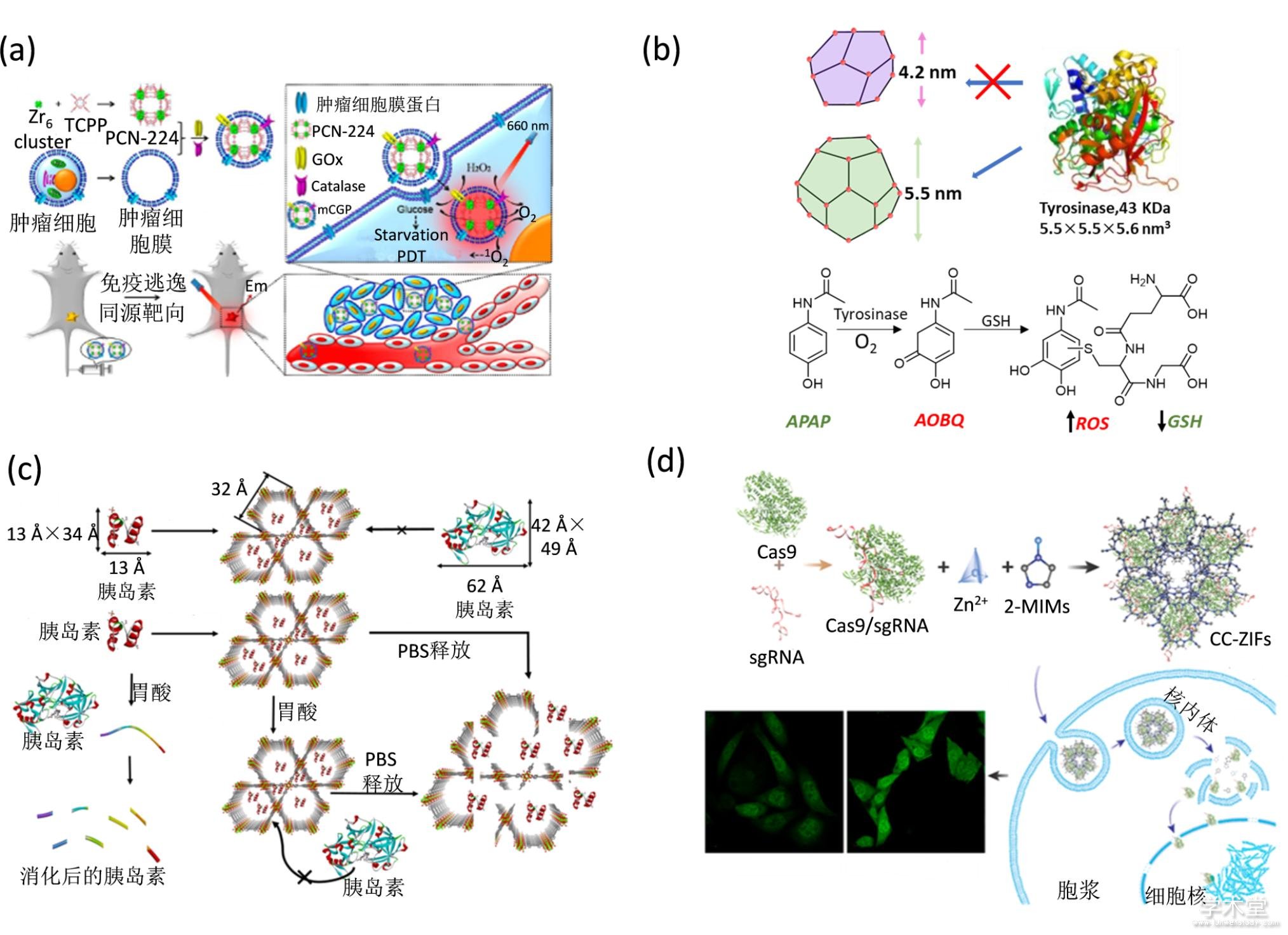
�����������л��Ǽܰ�����������ø����ʵ��ϸ���ڴ�˫��ˮ��Ϊ���ѧ�����ṩ������������������״̬[109,110] MOF�������������ø������������ϸ����������������ǣ����Ӫ����Դ��ʵ�ּ�������[111]��ͼ8a��. �ý����л��Ǽ�PCN-333�����Ұ���ø��������Ӧ����������λ��������Ϣʹǰҩ�����ɶ���ҩ�����GSH���ٺ�ROS�IJ�����ͼ8b�����Ӷ���Ч��ɱ������ϸ����������������[112]. �����л��Ǽܰ�����ҩ�����ø��organophosphorus acid anhydrolase��OPAA�����������ø�����ȶ����Լ���ʱ���ȶ��ԣ�ʵ�ָ�Чˮ�����Է���[113,114].
����һЩ�ڷ�������ҩ����ȵ��أ�������θ�����ȶ���������ٴ�Ӧ����Ч��Ȼ�д����. Ϊ�˽��������⣬Farha�������ý����л��Ǽ�NU-1000�����ȵ��أ��ȵ���@NU-1000�����Ӷ������ȵ�����θ�в�������[115]��ͼ8c��. ���Ƿ��֣��ȵ��صĸ�������30���Ӽ��ɴﵽ40wt%. NU-1000�����Ի����У�������pHΪ1�Ļ�������Ȼ�dz��ȶ�������ܹ������ȵ�����θ�ỷ���е��ȶ��ԣ��������ȵ��صĹ��ͺͻ���. NU-1000�����Ỻ��Һ��PBS�������н��⣬�ͷų��������ȵ��أ�ʹ�䷢�ӽ�Ѫ�ǵĹ���. �����ϵ���ܶ������β��ȶ������ܻᵼ�²����ȵ���й¶�Ӷ�ʧ����ǿ��Ծ���һ����������������ȵ��ص����������Ʊ��ڷ��ȵ����Ƽ��������źܴ��DZ��.
����Cas9��һ�ֻ���CRISPR��Clustered Regularly Interspersed Short Palindromic Repeats���ɴصĹ��ɼ���Ķ̻����ظ����У��Ļ���༭�������ף��������ڱ༭���鶯��ϸ���Ļ����飬�Ӷ�ʵ�ּ����Ļ�������[116]. Khashab������ZIF-8����Cas9�����ָ÷���������Ч�Ľ������ϸ��. ����ø������Ի����У�ZIF-8��Ϊ���屻���ӻ������⣬ʵ����ø�����ݺ��ͷ�Cas9��������Cas9���л���Ĺ���[117]��ͼ8d��. ����һ�ֽ����л��Ǽ�ZIF-90Ҳ������cas9���ף������������ϸ���ʻ�����. ϸ�����е�ATP������ZIF-90�Ľ���Ԫ��Zn2+��ǿ��λ���ã�ʹ��ZIF-90���⣬�Ӷ��ͷ�Cas9����[118]. Cas9������ϸ���еĻ�����У��ﵽ�����Ĭ��Ч��. ����������ϵ����ƺ��ʵļ���λ�㣬���Ը�������ϸ���Ļ����������ʺϳɣ��Ӷ���������ϸ���������Ҷ������ﵽ��������������Ŀ��.
������һ����������ҩ��——����Ҳ���㷺�����������ƣ�������С����RNA��siRNA����ͨ�������Ե���Ŀ��mRNA��������ŵ����ʵı���[119]. ͬ���أ�siRNA��Ҫ������ܽ��뵽ϸ���з��ӹ���. �����л��Ǽܾ�������ı������ʣ��ܹ��������ҩ��ͨ���������á�π-π����õȷ�ʽ���[120]. �����л��Ǽ�����siRNA֮����Խ�siRNA���䵽ϸ�����У����ϸ����ȡsiRNA��������ֹsiRNA��ˮ�⣬�ٽ�siRNA������ø�壬�Ӷ��ﵽ���õĻ�������Ч��[10]. DNAzyme�Ǿ��д����Ե�DNA���У����������Դ����Ǻ�������������Ǻ�����ѽ⣬��˳������ڻ�������[121]. ͬ���ģ�DNAzyme��Ҫ����������ܽ��뵽ϸ����. Wang������ZIF-8����DNAzyme����ﵽϸ����[88]��������ø�������ͷų�DNAzyme��ø����Zn2+��ʵ�ֻ�������.
����ͼ8 MOF�����ʸ��������ڼ�������
����Fig. 8 MOF-protein complex based therapy
����(a) �����л��Ǽ����ڼ�������[111]��(b) PCN-333�Ľṹʾ��ͼ��DZҩ����ʾ��ͼ[112]��(c) �����л��Ǽܰ����ȵ���ʾ��ͼ���ȵ����ͷ�ʾ��ͼ[115]��(d) Cas9@ZIF���Ʊ�ʾ��ͼ��ϸ������ʾ��ͼ�ͻ���༭Ч��[117]
����2.4�� ��������
�������ϻ��ڽ����л��Ǽܵ�����ҩ�������ϵ����ȡ����һ����չ[122]�����������ƵĹ�������Ȼ���ٵ�һ����Ч�������ߵ�����. ��ˣ�������Ч������������ϵ���зdz���Ҫ������.�����л��Ǽܾ��д�ıȱ�������ɵ��ijߴ硢�ɿصľ���ṹ�;���������Ե�����ͽ�����㣬��˷dz��ʺ�������������ƽ̨�Ĺ���. ����������ϵ�Ĺ�������ڵ�һ������ϵ��Ҫ����ϸ�µ���ƣ���Ҫ���ǵ������л��Ǽܵİ�������ͽ��⣬�Լ���ͬ�����ֶε�ʵʩ.
��������high-ZԪ�أ���Au[4]��Hf[5]��Bi[6] ��ϡ��Ԫ�ص�[123,124]�����������������л��Ǽܲ���. Liu���˽�������PDT��������ϵĽ����л��Ǽ�������ϵ. ���ǽ��Է��������еĽ�������Hf�������߲����������TCPP��װ�����������л��Ǽܣ���ͨ���������ξ��Ҷ����õ��������������ȶ��Ե�NMOF-PEG[64]��ͼ9a��. NMOF-PEG���зdz��߹��������������ڹ����£��ܹ���Ч�IJ�������̬��. High-ZԪ��Hf��Ϊ���������м���ͨ���������ӻ���������߷���Ч��. ��ˣ���������л��Ǽܼ��Ϲ��ѧ���ƺͷ��ƹ�����һ��. ͨ��β����ע�䣬NMOF-PEG����ͨ��EPRЧӦ��Ч�ĸ�����������λ. NMOF-PEG����Ƴ�������˽����л��Ǽܵ�����ͽ������Ŀɱ����Լ������������. ͨ��������ʵʩ�����ֶΣ������ѧ���ƺͷ����л���ϣ��ﵽ��Ч��������������Ч��.
�������˷��ƣ���������Ҳ����PDT���������������Ч��. ���������ǽ�������չ������һ�ָ�Ч�����������ֶΣ����ж��ڼ�������������Ƶ��о���Ϊ�㷺. ����������Ч�Ŀ�ԭ�ʵݺͲ���Ŀ��������ߵ�ԭ���������Ƶĵ�������Ч������. Lin���˽������Խ����л��Ǽܰ����������Ƽ���IDOi����������IDOi@TBC-Hf[5,10,15,20-tetra(pbenzoato)chlorin (H4TBC)]ϵͳ�����������������������ѧ���ƽ��������[125]����������˹����Խ����л��Ǽܵ�ҩ������ܣ�ͼ9b��. ��IDOi�����������л��ǼܵĿ��У�IDOi@TBC-Hf���Խ�IDOi�ͷŵ�����������IDOi����ѪҺѭ������ϵͳ��IDO����. ЭͬPDT�ͼ�������������ƿ��Դٽ�ԶλЧӦ������Զλ����������.
�������˹�����ƣ����õĹ����Ʒ������й������ƣ�Ҳ�ܹ���һ�����PDT������Ч��. ���磬Zhang���˽���߲����������Ϊ������������λ��װ�õ�Zr-FeP MOF. �ڽ���������£�Zr-FeP MOFͬʱ�����������ɻ��͵���̬����ʵ�ֹ������. Zr-FeP MOF�����Ը���siRNA���ڳ�Ĭ���ݿ˵��ף���Ч����ǿ���ѧ���Ƶ�Ч��. ���⣬��ͬһ�����������£�Zr-FeP MOF�Ĺ���ת��Ч��Ϊ33.7%��ʵ�е��¹�������. ͨ����������л��Ǽ���ϵ��ʵ���˹��ѧ���ơ��������ƺͻ��������������Ʒ���Эͬ����[126].
����Lee����ͨ��ԭλ�������ý����л��Ǽܰ���������graphitic carbon nitride (g-C3N4) nanosheets�Ϳװ�����װ�ػ���ҩ��Doxorubicin hydrochloride (DOX)���õ����ѧ���ƺͻ�������������ϵ[127]��ͼ9c��. ����ϵ�Ĺ�����Ҫ�����ڽ����л��Ǽܹ��������Ա��������ϵİ����ԡ��ɿ��Ժͽ����л��ǼܿDz�Ĵ��϶��. ͨ�����ƵĹ���������Yang�����ý����л��Ǽܰ�������ת�����Ͼ�������������PPy NPs���ͻ���ҩ����������, ʵ�ֻ��ƺ�����������[128]��ͼ9d��.���⣬���ý���߲����������Ϊ������װ�Ľ����л��Ǽܿ��Ը���ǰҩ���ӣ�����������[129]�ȣ�. �ڽ���������£������л��Ǽ�ʵ�ֹ������Ƶ�ͬʱ���为�ص������������Ȳ���һ�������������ڿ���������.
�����ܶ���֮����������Ч���ȵ�һ������Ч��Ҫ�úܶ�. ���ý����л��Ǽܹ�������������ϵ���Դ�������������Ч�����������ڽ��ת���������������������Ƶİ�֢.
����ͼ9�����л��Ǽ�������������
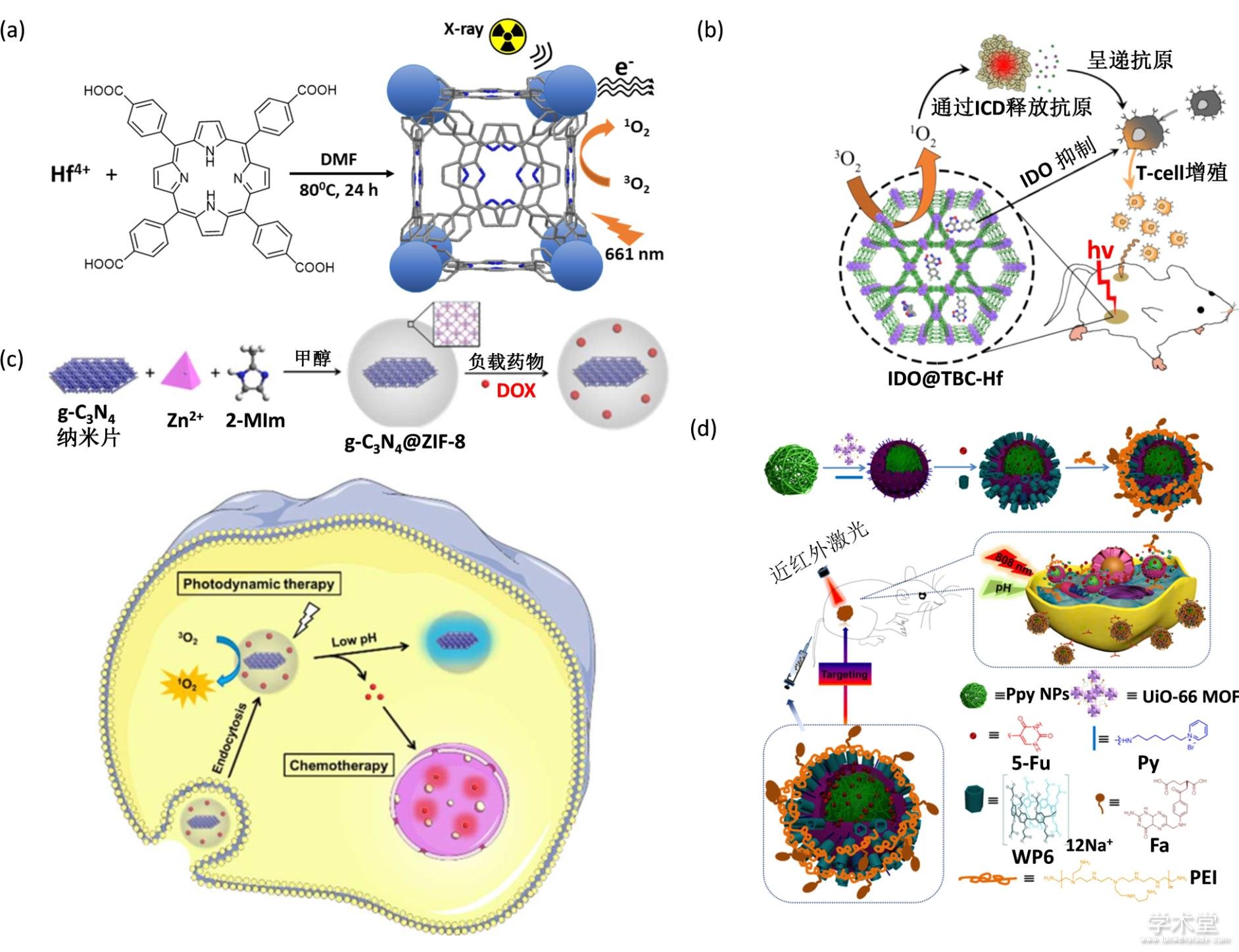
����Fig. 9 MOF for combined therapy
����(a) PDT�ͷ�����������[64]��(b) PDT��������������[125]��(c)���ƺ�PDT��������[127]��(d)���ƺ�����������[128]
����3 ���ܽ���չ��
������������������NMOF�����������о��ڽ�ʮ������ȡ���˽��Եijɹ�. ͨ���ǹ��۽������۽����ȷ����ܹ���Ч�ؽ�С����ҩ�︺���ڽ����л��Ǽ��ϣ�ͬʱ���ÿ�����ԭλ�����ȷ���Ҳ�ܹ�ʵ��MOF-��������ҩ��ĸ�Ч����. ֵ��һ����ǣ�ѡ�þ���ҩ����ԵĽ������Ļ��л����壬�����л��Ǽܱ���Ҳ�ܹ���Ƴɸ�Ч��ȫ��ҩ��. ����NMOF��ҩ�����ϵͳ�ڻ�ѧ���ơ����ѧ���ơ����������ƺ��������Ƶȼ������Ʒ�ʽ�Ͼ�������. ͨ������ҩ��С�������ڻ��ƻ��������ơ���װ���й����Ե�NMOF���ڹ��ѧ���ơ���װ���з��������ʵ�NMOF������ǿ���ơ�������������������ȷ����������в�ͬ���ܵ�NMOF������ϵ����չ�������õ�����Ч�����Խ����л��Ǽ�������ϵ�Ľ�һ��������Ӧ�����ŷdz��ش��ָ������. ���⣬���������ڽ����л��Ǽܵ�����������ϵ�����Dz��ѷ��֣����dz�����������������ϵ�Ĺ������Ӷ����ⵥһ����Ч�����Ѻ;��ж������õ�ȱ��.
�����ۺ������о������Dz��ѿ����������֬���壨liposome��[130,131]���ۺ�����������polymer��[132]����裨MSN��[133]������ʯīϩ��GO��[134]��ҩ�������ϵ��NMOF��Ϊҩ�������ϵ����һ�����ص�. ���ȣ�NMOF���нϸߵ�ҩ������Ч�ʣ��������ڰ�����������ʧ�����������. ��Σ������MSN��GO�������ϣ�NMOF���и��õ����������Ժ�����ɽ����ԣ��ܹ����������л���������ⳤ���ۻ������Ķ�������. ���ߣ�NMOF�ɷֿɵ������ڹ����ɿ�ҩ���ͷ����壬�����ڲ��������ε������ʵ�ֻ�����Դ�����ʣ�ATP�ȣ������ҩ���ͷ�. Ȼ����NMOFҲͬʱ����һЩȱ��. ���ȣ�NMOF��Ҫ��ͨ����λ������װ���ɣ����������ﻷ���е��ȶ��Խϲ�. ���磺ZIF-8������Fe��Zr����Ϊ�������Ľ����л��Ǽ�ͨ����PBS�����в��ȶ�. ��ˣ�ͨ����Ҫ���б������λ���������������������������ȶ���. ���⣬NMOFҩ�������ϵ�Ὣ�����������뵽�����ڣ����ܻ����һ���̶ȵĸ������������. ��ˣ�NMOF��ʵ��Ӧ����Ȼ����������Ҫ��������⣬�����������о���ս��������1��ʵ��ҩ��İ��������ɿ��ͷ�. Ŀǰ�������ϵ��ҩ��������л���һ���̶ȵ�ҩ��й¶�����ܻ�һ���̶��ϵ��¶������õij���. ͨ�������ı������Σ�����������塢Ҷ�ᡢ���������������ʶ����ӣ�����������ҩ��й¶��ͬʱ���ܸ����������������������������һ��DZ�ڵĽ������. ��2�����������������NMOF������. Ŀǰ���ֵĹ����Խ����л��Ǽ��ǻ���߲��������������PDT����������630 nm���ң���֯��������ޣ�һ��С��0.5 cm�������ںܴ�̶��������˹����Խ����л��Ǽܶ��������֯����������Ӧ��. ��ˣ���Ҫ�������������Ĺ������������ϳ����ͳ����������Խ����л��Ǽܣ���һ����ߺ�������л��Ǽܵ���������Ч��.��3��������ؽ����л��Ǽ����������ڵ��ȶ���. ���������֮���γ��ȶ�����λ����MOF�ṹ�γɵ�һ���ؼ�����������������������֮��Ҳ�����з��Ӽ�����������������. �����л��Ǽ��ڻ���ĸ��ӻ��������ֽ⣬�������ȱ�㣬Ҳ�����ŵ㣬�Ӿ���Ӧ�ö�������˸���Ӧ����Ҫ���ؽ����л��Ǽ����������ڵ��ȶ��Էdz���Ҫ. ��4�����NMOF-����������ȶ���������Ч��. ��Ȼ����ͨ����������������ȶ��Ժ�ˮ���ԣ����ǿ��ܻ�Ӱ�쵰�������ĽӴ����ߵ����ͷ�. ���⣬����ø��ɢ���뵽NMOF���еĻ����Բ���ȷ���˽�����������Ż�ø�������������������Ч��.
����NMOF������������������ײ��ϲ��߱����������ʣ��������������������Ҫ��Ӧ��DZ��. ����Ŀǰ�������������ѣ��������Ŷ�NMOFԽ��Խ������о�����Щ���ѻᱻ���ˣ��Ӷ�ʵ�������ĸ�Ч����ȫ�����õ�����Ч��.
���������
����[1] Idikio H A. Human cancer classification: a systems biology- based model integrating morphology, cancer stem cells, proteomics, and genomics. Journal of Cancer, 2011, 2: 107-115.
����[2] Heng H H, Stevens J B, Bremer S W, et al. Evolutionary mechanisms and diversity in cancer. Advances in Cancer Research, 2011, 112: 217-253.
����[3] Heng H H, Stevens J B, Bremer S W, et al. The evolutionary mechanism of cancer. Journal of Cellular Biochemistry, 2010, 109(6): 1072-1084.
����[4] Ulbrich K, Holá K, ?ubr V, et al. Targeted drug delivery with polymers and magnetic nanoparticles: covalent and noncovalent approaches, release control, and clinical studies. Chemical Reviews, 2016, 116(9): 5338-5431.
����[5] Kamaly N, Yameen B, Wu J, et al. Degradable controlled-release polymers and polymeric nanoparticles: mechanisms of controlling drug release. Chemical Reviews, 2016, 116(4): 2602-2663.
����[6] Zhou L, Qiu T, Lv F, et al. Self-assembled nanomedicines for anticancer and antibacterial applications. Advanced Healthcare Materials, 2018, 7(20): 1800670-1800698.
����[7] Giménez-Marqués M, Hidalgo T, Serre C, et al. Nanostructured metal–organic frameworks and their bio-related applications. Coordination Chemistry Reviews, 2016, 307: 342-360.
����[8] Horcajada P, Gref R, Baati T, et al. Metal-organic frameworks in biomedicine. Chemical Reviews, 2012, 112(2): 1232-1268.
����[9] Chowdhury M A. Metal-organic-frameworks for biomedical applications in drug delivery, and as MRI contrast agents. Journal of Biomedical Materials Research Part A, 2017, 105(4): 1184-1194.
����[10] He C, Lu K, Liu D, et al. Nanoscale metal-organic frameworks for the co-delivery of cisplatin and pooled siRNAs to enhance therapeutic efficacy in drug-resistant ovarian cancer cells. Journal of the American Chemical Society, 2014, 136(14): 5181-5184.
����[11] Guan Q, Li Y A, Li W Y, et al. Photodynamic therapy based on nanoscale metal-organic frameworks: from material design to cancer nanotherapeutics. Chemistry, an Asian Journal, 2018, 13(21): 3122-3149.
����[12] Ma T, Liu Y, Wu Q, et al. Quercetin-modified metal-organic frameworks for dual sensitization of radiotherapy in tumor tissues by inhibiting the carbonic anhydrase IX. ACS nano, 2019, 13(4): 4209-4219.
����[13] Lian X, Fang Y, Joseph E, et al. Enzyme-MOF (metal-organic framework) composites. Chemical Society Review, 2017, 46(11): 3386-3401.
����[14] Datt A, Ndiege N, Larsen S C. Development of porous nanomaterials for applications in drug delivery and imaging [M]. Nanomaterials for Biomedicine. American Chemical Society. 2012, 1119: 239-258.
����[15] Wu M-X, Yang Y-W. Metal–Organic Framework (MOF)-based drug/cargo delivery and cancer therapy. Advanced Materials, 2017, 29(23): 1606134-1606153.
����[16] Horcajada P, Serre C, Vallet-Regi M, et al. Metal-organic frameworks as efficient materials for drug delivery. Angewandte Chemie, 2006, 45(36): 5974-5978.
����[17] Gao X, Hai X, Baigude H, et al. Fabrication of functional hollow microspheres constructed from MOF shells: Promising drug delivery systems with high loading capacity and targeted transport. Scientific Reports, 2016, 6(1): 37705-37714.
����[18] Zhang F M, Dong H, Zhang X, et al. Postsynthetic modification of ZIF-90 for potential targeted codelivery of two anticancer drugs. ACS Applied Materials & Interfaces, 2017, 9(32): 27332-27337.
����[19] Fu J, Yu C, Li L, et al. Intracellular delivery of functional proteins and native drugs by cell-penetrating poly(disulfide)s. Journal of the American Chemical Society, 2015, 137(37): 12153-12160.
����[20] Bailey J B, Zhang L, Chiong J A, et al. Synthetic modularity of Protein-Metal-Organic Frameworks. Journal of the American Chemical Society, 2017, 139(24): 8160-8166.
����[21] Lian X, Erazo-Oliveras A, Pellois J P, et al. High efficiency and long-term intracellular activity of an enzymatic nanofactory based on metal-organic frameworks. Nature Communications, 2017, 8(1): 2075-2084.
����[22] Jung S, Kim Y, Kim S J, et al. Bio-functionalization of metal-organic frameworks by covalent protein conjugation. Chemical Communication (Camb), 2011, 47(10): 2904-2906.
����[23] Lian X, Chen Y P, Liu T F, et al. Coupling two enzymes into a tandem nanoreactor utilizing a hierarchically structured MOF. Chemical Science, 2016, 7(12): 6969-6973.
����[24] Cao Y, Wu Z, Wang T, et al. Immobilization of bacillus subtilis lipase on a Cu-BTC based hierarchically porous metal-organic framework material: a biocatalyst for esterification. Dalton transactions, 2016, 45(16): 6998-7003.
����[25] Liu W L, Yang N S, Chen Y T, et al. Lipase-supported metal-organic framework bioreactor catalyzes warfarin synthesis. Chemistry, 2015, 21(1): 115-119.
����[26] Patra S, Hidalgo Crespo T, Permyakova A, et al. Design of metal organic framework–enzyme based bioelectrodes as a novel and highly sensitive biosensing platform. Journal of Materials Chemistry B, 2015, 3(46): 8983-8992.
����[27] Fracaroli A M, Siman P, Nagib D A, et al. Seven Post-synthetic covalent reactions in tandem leading to enzyme-like complexity within Metal-Organic Framework crystals. Journal of the American Chemical Society, 2016, 138(27): 8352-8355.
����[28] Liang W, Ricco R, Maddigan N K, et al. Control of structure topology and spatial distribution of biomacromolecules in protein@ZIF-8 biocomposites. Chemistry of Materials, 2018, 30(3): 1069-1077.
����[29] Sontz P A, Bailey J B, Ahn S, et al. A Metal Organic Framework with spherical protein nodes: rational chemical design of 3D protein crystals. Journal of the American Chemical Society, 2015, 137(36): 11598-11601.
����[30] Wang Z, Hu S, Yang J, et al. Nanoscale Zr-based MOFs with tailorable size and introduced mesopore for protein delivery. Advanced Functional Materials, 2018, 28(16): 1707356-1707364.
����[31] Zhang H, Lv Y, Tan T, et al. Atomistic simulation of protein encapsulation in metal-organic frameworks. The Journal of Physical Chemistry B, 2016, 120(3): 477-484.
����[32] Larsen R W, Wojtas L, Perman J, et al. Mimicking heme enzymes in the solid state: metal-organic materials with selectively encapsulated heme. Journal of the American Chemical Society, 2011, 133(27): 10356-10359.
����[33] Liu X, Qi W, Wang Y, et al. A facile strategy for enzyme immobilization with highly stable hierarchically porous metal–organic frameworks. Nanoscale, 2017, 9(44): 17561-17570.
����[34] Wang S, Chen Y, Wang S, et al. DNA-functionalized metal-organic framework nanoparticles for intracellular delivery of proteins. Journal of the American Chemical Society, 2019, 141(6): 2215-2219.
����[35] Lykourinou V, Chen Y, Wang X S, et al. Immobilization of MP-11 into a mesoporous metal-organic framework, MP-11@mesoMOF: a new platform for enzymatic catalysis. Journal of the American Chemical Society, 2011, 133(27): 10382-10385.
����[36] Chen Y, Lykourinou V, Vetromile C, et al. How can proteins enter the interior of a MOF? Investigation of cytochrome c translocation into a MOF consisting of mesoporous cages with microporous windows. Journal of the American Chemical Society, 2012, 134(32): 13188-13191.
����[37] Feng D, Liu T F, Su J, et al. Stable metal-organic frameworks containing single-molecule traps for enzyme encapsulation. Nature Communications, 2015, 6: 5979-5986.
����[38] Lian X, Chen Y-P, Liu T-F, et al. Coupling two enzymes into a tandem nanoreactor utilizing a hierarchically structured MOF. Chemical Science, 2016, 7(12): 6969-6973.
����[39] Zhu G, Zhang M, Bu Y, et al. Enzyme-embedded metal-organic framework colloidosomes via an emulsion-based approach. Chemistry, an Asian journal, 2018, 13(19): 2891-2896.
����[40] Nadar S S, Rathod V K. Facile synthesis of glucoamylase embedded metal-organic frameworks (glucoamylase-MOF) with enhanced stability. International Journal of Biological Macromolecules, 2017, 95: 511-519.
����[41] Salgaonkar M, Nadar S S, Rathod V K. Combi-metal organic framework (Combi-MOF) of alpha-amylase and glucoamylase for one pot starch hydrolysis. International Journal of Biological Macromolecules, 2018, 113: 464-475.
����[42] Lyu F, Zhang Y, Zare R N, et al. One-pot synthesis of protein-embedded metal-organic frameworks with enhanced biological activities. Nano Letters, 2014, 14(10): 5761-5765.
����[43] Liao F S, Lo W S, Hsu Y S, et al. Shielding against unfolding by embedding enzymes in metal-organic frameworks via a de novo approach. Journal of the American Chemical Society, 2017, 139(19): 6530-6533.
����[44] Liang K, Ricco R, Doherty C M, et al. Biomimetic mineralization of metal-organic frameworks as protective coatings for biomacromolecules. Nature Communications, 2015, 6: 7240-7247.
����[45] Maddigan N K, Tarzia A, Huang D M, et al. Protein surface functionalisation as a general strategy for facilitating biomimetic mineralisation of ZIF-8. Chemical Science, 2018, 9(18): 4217-4223.
����[46] Chen G, Huang S, Kou X, et al. A convenient and versatile amino-acid-boosted biomimetic strategy for the nondestructive encapsulation of biomacromolecules within metal-organic frameworks. Angewandte Chemie, 2019, 58(5): 1463-1467.
����[47] Liang W, Xu H, Carraro F, et al. Enhanced activity of enzymes encapsulated in hydrophilic metal-organic frameworks. Journal of the American Chemical Society, 2019, 141(6): 2348-2355.
����[48] Imaz I, Rubio-Martinez M, An J, et al. Metal-biomolecule frameworks (MBioFs). Chem Commun (Camb), 2011, 47(26): 7287-7302.
����[49] An J, Farha O K, Hupp J T, et al. Metal-adeninate vertices for the construction of an exceptionally porous metal-organic framework. Nature Communications, 2012, 3: 604-609.
����[50] An J, Geib S J, Rosi N L. Cation-triggered drug release from a porous zinc?adeninate metal?organic framework. Journal of the American Chemical Society, 2009, 131(24): 8376-8377.
����[51] An J, Shade C M, Chengelis-Czegan D A, et al. Zinc-adeninate metal-organic framework for aqueous encapsulation and sensitization of near-infrared and visible emitting lanthanide cations. Journal of the American Chemical Society, 2011, 133(5): 1220-1223.
����[52] Zhang L, Lei J, Ma F, et al. A porphyrin photosensitized metal–organic framework for cancer cell apoptosis and caspase responsive theranostics. Chemical Communications, 2015, 51(54): 10831-10834.
����[53] Park J, Jiang Q, Feng D, et al. Size-controlled synthesis of porphyrinic metal-organic framework and functionalization for targeted photodynamic therapy. Journal of the American Chemical Society, 2016, 138(10): 3518-3525.
����[54] Wang W, Wang L, Li Z, et al. BODIPY-containing nanoscale metal-organic frameworks for photodynamic therapy. Chem Commun (Camb), 2016, 52(31): 5402-5405.
����[55] Chen J, Zhang X, Huang C, et al. Osteogenic activity and antibacterial effect of porous titanium modified with metal-organic framework films. Journal of Biomedical Materials Research Part A, 2017, 105(3): 834-846.
����[56] Seyedpour S F, Rahimpour A, Najafpour G. Facile in-situ assembly of silver-based MOFs to surface functionalization of TFC membrane: A novel approach toward long-lasting biofouling mitigation. Journal of Membrane Science, 2019, 573: 257-269.
����[57] Wu Y, Luo Y, Zhou B, et al. Porous metal-organic framework (MOF) Carrier for incorporation of volatile antimicrobial essential oil. Food Control, 2019, 98: 174-178.
����[58] Guo X, Guo B, Wang Y, et al. Preparation of spherical metal-organic frameworks encapsulating ag nanoparticles and study on its antibacterial activity. Materials Science & Engineering C, Materials for Biological Applications, 2017, 80: 698-707.
����[59] Lan G, Ni K, Veroneau S S, et al. Nanoscale metal-organic framework hierarchically combines high-Z components for multifarious radio-enhancement. Journal of the American Chemical Society, 2019, 141(17): 6859-6863.
����[60] Chen Z -F, Xue H-B, Wu L-R, et al. Bifunctional (3,6)-connected Gd(III) metal–organic framework material with catalytic properties for the cyanosilylation reaction and anti-gastric cancer activity. Journal of Cluster Science, 2018, 29(6): 1269-1274.
����[61] Qi X, Tian H, Dang X, et al. A bimetallic Co/Mn metal–organic-framework with a synergistic catalytic effect as peroxidase for the colorimetric detection of H2O2. Analytical Methods, 2019, 11(8): 1111-1124.
����[62] Sun L-L, Li Y-H, Shi H. A Ketone Functionalized Gd(III)-MOF with Low Cytotoxicity for Anti-Cancer Drug Delivery and Inhibiting Human Liver Cancer Cells. Journal of Cluster Science, 2018, 30(1): 251-258.
����[63] Wan S S, Cheng Q, Zeng X, et al. A Mn(III)-Sealed Metal-Organic Framework Nanosystem for Redox-Unlocked Tumor Theranostics. ACS nano, 2019, 13(6): 6561-6571.
����[64] Prencipe G, Tabakman S M, Welsher K, et al. PEG Branched Polymer for Functionalization of Nanomaterials with Ultralong Blood Circulation. Journal of the American Chemical Society, 2009, 131(13): 4783-4787.
����[65] Liu J, Yang Y, Zhu W, et al. Nanoscale metal-organic frameworks for combined photodynamic & radiation therapy in cancer treatment. Biomaterials, 2016, 97: 1-9.
����[66] Abanades Lazaro I, Haddad S, Sacca S, et al. Selective Surface PEGylation of UiO-66 Nanoparticles for Enhanced Stability, Cell Uptake, and pH-Responsive Drug Delivery. Chem, 2017, 2(4): 561-578.
����[67] Chen T T, Yi J T, Zhao Y Y, et al. Biomineralized Metal-Organic Framework Nanoparticles Enable Intracellular Delivery and Endo-Lysosomal Release of Native Active Proteins. Journal of the American Chemical Society. 2018, 140(31): 9912-9920.
����[68] Cheng G, Li W, Ha L, et al. Self-Assembly of Extracellular Vesicle-like Metal-Organic Framework Nanoparticles for Protection and Intracellular Delivery of Biofunctional Proteins. Journal of the American Chemical Society, 2018, 140(23): 7282-7291.
����[69] Zhang L, Wang Z, Zhang Y, et al. Erythrocyte Membrane Cloaked Metal-Organic Framework Nanoparticle as Biomimetic Nanoreactor for Starvation-Activated Colon Cancer Therapy. ACS nano, 2018, 12(10): 10201-10211.
����[70] Wan X, Zhong H, Pan W, et al. Programmed Release of Dihydroartemisinin for Synergistic Cancer Therapy Using a CaCO3 Mineralized Metal-Organic Framework. Angewandte Chemie, 2019, 58(40): 14134-14139.
����[71] Azizi Vahed T, Naimi-Jamal M R, Panahi L. Alginate-coated ZIF-8 metal-organic framework as a green and bioactive platform for controlled drug release. Journal of Drug Delivery Science and Technology, 2019, 49: 570-576.
����[72] Ren M, Li H, Liu H, et al. A Biocompatible GdIII–Organic Framework Incorporating Polar Pores for pH-Sensitive Anti-Cancer Drug Delivery and Inhibiting Human Bone Tumour Cells. Australian Journal of Chemistry, 2019, 72(3): 233.
����[73] Gupta V, Tyagi S, Paul A K. Development of Biocompatible Iron-Carboxylate Metal Organic Frameworks for pH-Responsive Drug Delivery Application. Journal of Nanoscience and Nanotechnology, 2019, 19(2): 646-654.
����[74] Zhao L-C, Tang M, Zhang Q-H, et al. Fabrication of a Porous Metal-Organic Framework with Polar Channels for 5-Fu Delivery and Inhibiting Human Osteosarcoma Cells. Journal of Chemistry, 2018, 2018: 1-7.
����[75] Cai Y, Sheng Z, Wang J. A Biocompatible Zinc(II)-based Metal-organic Framework for pH Responsive Drug Delivery and Anti-Lung Cancer Activity. Zeitschrift Für Anorganische und Allgemeine Chemie, 2018, 644(16): 877-882.
����[76] Du X, Fan R, Qiang L, et al. Controlled Zn2+-Triggered Drug Release by Preferred Coordination of Open Active Sites within Functionalization Indium Metal Organic Frameworks. ACS Applied Materials & Interfaces, 2017, 9(34): 28939-28948.
����[77] Luo Z, Jiang L, Yang S, et al. Light-Induced Redox-Responsive Smart Drug Delivery System by Using Selenium-Containing Polymer@MOF Shell/Core Nanocomposite. Advanced Healthcare Materials, 2019, e1900406.
����[78] Javanbakht S, Pooresmaeil M, Namazi H. Green one-pot synthesis of carboxymethylcellulose/Zn-based metal-organic framework/graphene oxide bio-nanocomposite as a nanocarrier for drug delivery system. Carbohydrate Polymers, 2019, 208: 294-301.
����[79] Xue Z, Zhu M, Dong Y, et al. An integrated targeting drug delivery system based on the hybridization of graphdiyne and MOFs for visualized cancer therapy. Nanoscale, 2019, 11, 11709.
����[80] Dong K, Zhang Y, Zhang L, et al. Facile preparation of metal-organic frameworks-based hydrophobic anticancer drug delivery nanoplatform for targeted and enhanced cancer treatment. Talanta, 2019, 194: 703-708.
����[81] Zhang W, Guo T, Wang C, et al. MOF Capacitates Cyclodextrin to Mega-Load Mode for High-Efficient Delivery of Valsartan. Pharmaceutical research, 2019, 36(8): 117-129.
����[82] Nasihat Sheno N, Farhadi S, Maleki A, et al. A novel approach for the synthesis of phospholipid bilayer-coated zeolitic imidazolate frameworks: preparation and characterization as a pH-responsive drug delivery system. New Journal of Chemistry, 2019, 43(4): 1956-1963.
����[83] Chen Z-X, Liu M-D, Zhang M-K, et al. Interfering with Lactate-Fueled Respiration for Enhanced Photodynamic Tumor Therapy by a Porphyrinic MOF Nanoplatform. Advanced Functional Materials, 2018, 28(36): 1803498-1803509.
����[84] Gao S, Zheng P, Li Z, et al. Biomimetic O2-Evolving metal-organic framework nanoplatform for highly efficient photodynamic therapy against hypoxic tumor. Biomaterials, 2018, 178: 83-94.
����[85] He L, Brasino M, Mao C, et al. DNA-assembled core-satellite upconverting-metal-organic framework nanoparticle superstructures for efficient photodynamic therapy. Small, 2017, 13(24): 1700504-1700510.
����[86] Kan J L, Jiang Y, Xue A, et al. Surface Decorated Porphyrinic Nanoscale Metal-Organic Framework for Photodynamic Therapy. Inorganic Chemistry, 2018, 57(9): 5420-5428.
����[87] Fan W, Huang P, Chen X. Overcoming the Achilles' heel of photodynamic therapy. Chemical Society Reviews, 2016, 45(23): 6488-6519.
����[88] Shen Y, Shuhendler A J, Ye D, et al. Two-photon excitation nanoparticles for photodynamic therapy. Chemical Society Reviews, 2016, 45(24): 6725-6741.
����[89] Li Y-A, Zhao X-D, Yin H-P, et al. A drug-loaded nanoscale metal–organic framework with a tumor targeting agent for highly effective hepatoma therapy. Chemical Communications, 2016, 52(98): 14113-14116.
����[90] Lismont M, Dreesen L, Wuttke S. Metal-Organic Framework nanoparticles in photodynamic therapy: current Status and perspectives. Advanced Functional Materials, 2017, 27(14): 1606314-1606329.
����[91] Min H, Wang J, Qi Y, et al. Biomimetic metal-organic framework nanoparticles for cooperative combination of antiangiogenesis and photodynamic therapy for enhanced efficacy. Advanced Materials, 2019, 31(15): 1808200-1808210.
����[92] Park J, Feng D, Yuan S, et al. Photochromic metal-organic frameworks: reversible control of singlet oxygen generation. Angewandte Chemie, 2015, 54(2): 430-435.
����[93] Wang D, Wu H, Lim W Q, et al. A mesoporous nanoenzyme derived from metal-organic frameworks with endogenous oxygen generation to alleviate tumor hypoxia for significantly enhanced photodynamic therapy. Advanced Materials, 2019, 1901893-1901901.
����[94] Wang H, Chen Y, Wang H, et al. DNAzyme-loaded metal–organic frameworks (MOFs) for self-sufficient gene therapy. Angewandte Chemie International Edition, 2019, 58(22): 7380-7384.
����[95] Lu K, He C, Lin W. Nanoscale metal-organic framework for highly effective photodynamic therapy of resistant head and neck cancer. Journal of the American Chemical Society, 2014, 136(48): 16712-16715.
����[96] Lu K, He C, Lin W. A chlorin-based nanoscale metal-organic framework for photodynamic therapy of colon cancers. Journal of the American Chemical Society, 2015, 137(24): 7600-7603.
����[97] Park J, Jiang Q, Feng D, et al. Controlled generation of singlet oxygen in living cells with tunable ratios of the photochromic switch in metal–organic frameworks. Angewandte Chemie International Edition, 2016, 55(25): 7188-7193.
����[98] Ma Y, Li X, Li A, et al. H2S-Activable MOF nanoparticle photosensitizer for effective photodynamic therapy against cancer with controllable singlet-oxygen release. Angewandte Chemie International Edition, 2017, 56(44): 13752-13756.
����[99] Zhang W, Lu J, Gao X, et al. Enhanced photodynamic therapy by reduced levels of intracellular glutathione obtained by employing a nano-MOF with CuII as the active center. Angewandte Chemie International Edition, 2018, 57(18): 4891-4896.
����[100] Bim Júnior O, Bedran-Russo A, Flor J B S, et al. Encapsulation of collagenase within biomimetically mineralized metal–organic frameworks: designing biocomposites to prevent collagen degradation. New Journal of Chemistry, 2019, 43(2): 1017-1024.
����[101] Gkaniatsou E, Sicard C, Ricoux R, et al. Enzyme encapsulation in mesoporous metal-organic frameworks for selective biodegradation of harmful dye molecules. Angewandte Chemie, 2018, 57(49): 16141-16146.
����[102] Kato S, Otake K I, Chen H, et al. Zirconium-based metal-organic frameworks for the removal of protein-bound uremic toxin from human serum albumin. Journal of the American Chemical Society, 2019, 141(6): 2568-2576.
����[103] Liang Z, Yang Z, Yuan H, et al. A protein@metal-organic framework nanocomposite for pH-triggered anticancer drug delivery. Dalton transactions, 2018, 47(30): 10223-10228.
����[104] Luzuriaga M A, Welch R P, Dharmarwardana M, et al. Enhanced stability and controlled delivery of MOF-encapsulated vaccines and their immunogenic response in vivo. ACS Applied Materials & Interfaces, 2019, 11(10): 9740-9746.
����[105] Nath I, Chakraborty J, Verpoort F. Metal organic frameworks mimicking natural enzymes: a structural and functional analogy. Chemical Society Reviews, 2016, 45(15): 4127-4170.
����[106] Shieh F K, Wang S C, Yen C I, et al. Imparting functionality to biocatalysts via embedding enzymes into nanoporous materials by a de novo approach: size-selective sheltering of catalase in metal-organic framework microcrystals. Journal of the American Chemical Society, 2015, 137(13): 4276-4279.
����[107] Xu M, Yuan S, Chen X Y, et al. Two-dimensional metal-organic framework nanosheets as an enzyme inhibitor: modulation of the alpha-chymotrypsin activity. Journal of the American Chemical Society, 2017, 139(24): 8312-8319.
����[108] Yu M, You D, Zhuang J, et al. Controlled release of naringin in metal-organic framework-loaded mineralized collagen coating to simultaneously enhance osseointegration and antibacterial activity. ACS Applied Materials & Interfaces, 2017, 9(23): 19698-19705.
����[109] Cheng H, Zhu J-Y, Li S-Y, et al. An O2 self-sufficient biomimetic nanoplatform for highly specific and efficient photodynamic therapy. Advanced Functional Materials, 2016, 26(43): 7847-7860.
����[110] Liu J, Liu T, Du P, et al. Metal-Organic Framework (MOF) hybrid as a tandem catalyst for enhanced therapy against hypoxic tumor cells. Angewandte Chemie, 2019, 58(23): 7808-7812.
����[111] Li S Y, Cheng H, Xie B R, et al. Cancer cell membrane camouflaged cascade bioreactor for cancer targeted starvation and photodynamic therapy. ACS nano, 2017, 11(7): 7006-7018.
����[112] Lian X, Huang Y, Zhu Y, et al. Enzyme-MOF nanoreactor activates nontoxic paracetamol for cancer therapy. Angewandte Chemie International Edition, 2018, 57(20): 5725-5730.
����[113] Li P, Moon S Y, Guelta M A, et al. Encapsulation of a nerve agent detoxifying enzyme by a mesoporous zirconium metal-organic framework engenders thermal and long-term stability. Journal of the American Chemical Society, 2016, 138(26): 8052-8055.
����[114] Li P, Moon S Y, Guelta M A, et al. Nanosizing a metal-organic framework enzyme carrier for accelerating nerve agent hydrolysis. ACS nano, 2016, 10(10): 9174-9182.
����[115] Chen Y, Li P, Modica J A, et al. Acid-resistant mesoporous metal-organic framework toward oral insulin delivery: protein encapsulation, protection, and release. Journal of the American Chemical Society, 2018, 140(17): 5678-5681.
����[116] Siu K H, Chen W. Riboregulated toehold-gated gRNA for programmable CRISPR-Cas9 function. Nature Chemical Biology, 2019, 15(3): 217-220.
����[117] Alsaiari S K, Patil S, Alyami M, et al. Endosomal escape and delivery of CRISPR/Cas9 genome editing machinery enabled by nanoscale zeolitic imidazolate framework. Journal of the American Chemical Society, 2018, 140(1): 143-146.
����[118] Yang X, Tang Q, Jiang Y, et al. Nanoscale ATP-responsive zeolitic imidazole framework-90 as a general platform for cytosolic protein delivery and genome editing. Journal of the American Chemical Society, 2019, 141(9): 3782-3786.
����[119] Ngamcherdtrakul W, Yantasee W. siRNA therapeutics for breast cancer: recent efforts in targeting metastasis, drug resistance, and immune evasion. Translational Research : the Journal of Laboratory and Clinical Medicine, 2019, 214: 105-120.
����[120] Zhu W, Guo J, Agola J O, et al. Metal-organic framework nanoparticle-assisted cryopreservation of red blood cells. Journal of the American Chemical Society, 2019, 141(19): 7789-7796.
����[121] Yin C, Ye B, Tan W, Wang H, et al. An allosteric dual-DNAzyme unimolecular probe for colorimetric detection of copper(II). Journal of the American Chemical Society, 2009, 131:14624–14625.
����[122] Rojas S, Arenas-Vivo A, Horcajada P. Metal-organic frameworks: A novel platform for combined advanced therapies. Coordination Chemistry Reviews, 2019, 388: 202-226.
����[123] Ni K, Lan G, Veroneau S S, et al. Nanoscale metal-organic frameworks for mitochondria-targeted radiotherapy-radiodynamic therapy. Nature Communications, 2018, 9(1): 4321-4333.
����[124] Chao Y, Liang C, Yang Y, et al. Highly effective radioisotope cancer therapy with a non-therapeutic isotope delivered and sensitized by nanoscale coordination polymers. ACS nano, 2018, 12(8): 7519-7528.
����[125] Lu K, He C, Guo N, et al. Chlorin-based nanoscale metal-organic framework systemically rejects colorectal cancers via synergistic photodynamic therapy and checkpoint blockade immunotherapy. Journal of the American Chemical Society, 2016, 138(38): 12502-12510.
����[126] Zhang K, Meng X, Cao Y, et al. Metal-organic framework nanoshuttle for synergistic photodynamic and low-temperature photothermal therapy. Advanced Functional Materials, 2018, 28(42): 1804634-1804643.
����[127] Chen R, Zhang J, Wang Y, et al. Graphitic carbon nitride nanosheet@metal–organic framework core–shell nanoparticles for photo-chemo combination therapy. Nanoscale, 2015, 7(41): 17299-17305.
����[128] Wu M X, Yan H J, Gao J, et al. Multifunctional supramolecular materials constructed from polypyrrole@UiO-66 nanohybrids and pillararene nanovalves for targeted chemophotothermal therapy. ACS Applied Materials & Interfaces, 2018, 10(40): 34655-34663.
����[129] Zhang H, Tian X T, Shang Y, et al. Theranostic Mn-porphyrin metal-organic frameworks for magnetic resonance imaging-guided nitric oxide and photothermal synergistic therapy. ACS Applied Materials & Interfaces, 2018, 10(34): 28390-28398.
����[130] Liu X, Situ A, Kang Y, et al. Irinotecan delivery by lipid-coated mesoporous silica nanoparticles shows improved efficacy and safety over liposomes for pancreatic cancer. ACS nano, 2016, 10(2): 2702-2715.
����[131] Zhang Y, Chan H F, Leong K W. Advanced materials and processing for drug delivery: the past and the future. Advanced Drug Delivery Reviews, 2013, 65(1): 104-120.
����[132] Li F, Zhao X, Wang H, et al. Multiple layer-by-layer lipid-polymer hybrid nanoparticles for improved FOLFIRINOX chemotherapy in pancreatic tumor models. Advanced Functional Materials, 2015, 25(5): 788-798.
����[133] Lijue Chen X S, Taowang, Li He, Sarah Shigdar, Wei Duanb and Lingxue Kong. Overcoming acquired drug resistance in colorectal cancer cells by targeted delivery of 5-FU with EGF grafted hollow mesoporous silica nanoparticles. Nanoscale, 2015, 7:14080-14092.
����[134] Fan X, Jiao G, Zhao W, et al. Magnetic Fe3O4-graphene composites as targeted drug nanocarriers for pH-activated release. Nanoscale, 2013, 5(3): 1143-1152.


