摘要:肩袖钙化性肌腱炎(rotator cuff calcific tendinitis, RCCT)是导致肩关节疼痛常见的原因之一,多发于30~60岁的女性人群。急性发作期肩痛剧烈,但经过有效的治疗症状会逐渐缓解而治愈。作为一种自限性疾病,RCCT的病理过程、辅助检查、治疗方式及其时机在临床上仍存在一些误区。本文就RCCT的诊疗误区与对策进行概述。
关键词:肩袖钙化性肌腱炎;诊疗误区;对策;病例报告;综述;
Abstract:Rotator cuff calcific tendinitis(RCCT) is a common cause of shoulder pain, usually occurs in women aged between 30 and60 years. Although patients present severe pain during acute phase, the symptom could be released by effective treatments. Because of a self-healing disease, RCCT presents certain pitfalls in terms of pathogenesis, imaging diagnosis, choice and timing of treatments. This article reported a case of RCCT and reviewed the pitfalls and strategies focused on diagnosis and treatment.
Keyword:rotator cuff calcific tendinitis; diagnostic and treatment pitfalls; strategy; case report; review;
肩袖钙化性肌腱炎(rotator cuff calcific tendinitis,RCCT)占肩关节疼痛原因的5%~40%,好发于30~60岁的女性人群,冈上肌腱约占80%,冈下肌腱占15%,肩胛下肌腱占5%[1,2,3,4,5]。约50%的患者无明显诱因,其病因尚不完全清楚,可能与退变、创伤、缺氧、内分泌代谢紊乱等有关[6,7,8,9,10]。由于羟基磷灰石[Ca10(PO4)6(OH)2]沉积,炎性刺激导致突发性、刀割样剧烈疼痛和肩关节活动受限[11]。
RCCT的病理变化有多种学说[9,10,12]。Uhthoff等[6]提出的反应性钙化理论得到普遍认可,把RCCT分为3个阶段:钙化前期、钙化期以及钙化后期。钙化期又分形成期、休眠期和吸收期。其中吸收期又称急性发作期,顾名思义,此时钙化灶吸收,巨噬细胞和多核巨细胞活跃,发生较为剧烈的炎症反应,此期疼痛最为剧烈[3,6,13]。RCCT是自限性疾病,钙化病灶有可能自行吸收痊愈。
RCCT的诊断需要与肩袖肌腱损伤、肩峰下撞击综合征、颈椎病、肱二头肌腱炎、痛风性关节炎等疾病相鉴别,特别要同退行性或营养不良性钙化相鉴别,后两者的好发人群年龄大,病灶部位是腱-骨相连接处,不属于自限性疾病[6,14]。吸收期钙化灶累及肩峰下滑囊、肱二头肌长头腱或肱骨大结节骨组织,也可累及至肱二头肌腱短头等部位[3,15,16]。
关于RCCT和肩袖肌腱损伤是否具有相关性目前存在争议。Brinkman等[2]发现云雾状钙化灶和肩袖损伤有显着相关性,但无法通过钙化灶的部位或体积预测是否存在肩袖损伤。Beckmann等[14]通过回顾性研究发现RCCT组和肩痛对照组的肩袖损伤患病率无统计学差异,认为RCCT和肩袖损伤出自于2个不同的病理过程。
影像学检查对诊断与鉴别诊断具有重要价值,X线检查具有快捷、方便和价廉等优点,是RCCT的首选影像学检查。典型的病例可见距离肩袖肌腱肱骨附着处1.5~2 cm的高信号钙化灶。目前普遍采用G?rt?ner等[17]提出的影像学分型对钙化灶描述:Ⅰ型:钙化灶密度高且分界明显;Ⅱ型:钙化灶密度高但分界不明显,或者分界明显但钙化灶密度低;Ⅲ型:钙化灶密度低且分界不明显呈云雾状。CT可以更加清晰地显示钙化灶,定位钙化灶的准确位置,尤其可以明确发现易漏诊的肩胛下肌腱的钙化灶,应作为RCCT手术治疗或非手术治疗前的进一步检查。超声(ultrasound,US)检查可以有效探及钙化病灶,明确钙化灶的位置,观察钙化灶的体积、质地、形状且无辐射,可用于孕妇等人群,但对检查者的经验和技术依赖性较高[4,18]。MRI有助于鉴别诊断和发现肩袖的伴随损伤,但通常检查时间较长且费用较高[2,19]。
临床医师只有深刻掌握RCCT的流行病学、病理过程、辅助检查等基本知识,才能正确选择治疗方式及其手术治疗时机。本文报告1例手术治疗RCCT患者,并结合文献对其治疗展开综述。
1 病例报告
患者,男,57岁,右肩无诱因阵发性剧痛3 d余,疼痛呈针刺、刀割样,严重影响睡眠和生活质量。查体:右肩呈被动体位,左手托举右肘。右肩峰前下部压痛,肩关节活动受限。右肩关节X线示:肩峰下高密度片状阴影(图1a),临床诊断钙化性冈上肌腱炎。
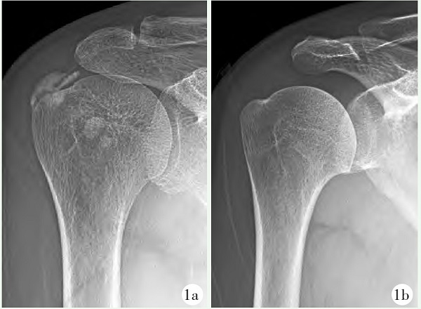
图1 患者,男,57岁
1a:术前X线片示肩峰下高密度阴影1b:保守治疗5周后,镜下未发现病灶,术后右肩关节X线片示肩峰下高密度片状阴影完全消失
患者强烈要求住院手术治疗,但因床位紧张,医师给予消炎镇痛药和理疗,先行保守治疗,回家待床手术治疗。患者5周后入院,疼痛较前有所缓解,但仍影响肩关节部分功能,完成术前常规检查后,全麻下行肩关节镜探查。术中反复探查肩峰下间隙与盂肱关节,均未发现钙化病灶并且肩袖完好。术中进行X线片透视检查也未发现钙化灶,于是射频清理增生的炎性滑膜结束手术。术后右肩关节X线片检查示:肩峰下高密度片状阴影完全消失(图1b)。
2 讨论与综述
该例肩关节钙化性肌腱炎的诊疗中有如下误区:患者自发病到手术间隔时间5周之久,正处于炎症反应吸收期,疼痛剧烈,经过药物和理疗等保守治疗后,钙化病灶是可以自行吸收的,忽视了病情有自愈的可能性。术前没有进行病情检查和严格的手术适应证评估,术前进行近期的影像学复查是十分必要的,只根据5周前X线检查直接采取手术治疗,结果术中未能发现病灶。
2.1 非手术治疗
对于RCCT优先采取保守治疗,包括适当休息、冰敷、物理治疗、应用非甾体类抗炎药(nonsteroi?dal anti-inflammatory drugs,NSAIDs)等。应用NSAIDs被认为是RCCT首选的治疗方式,可以有效缓解大多数肩痛症状[20]。有创的非手术治疗适用于症状严重或迁延不愈的RCCT患者,其中肩峰下糖皮质激素注射(subacromial corticosteroid injection,SAI)费用较少、并发症发病率低,是临床最常应用的治疗方式[6,21]。de Witte等[22]发现73%的RCCT经SAI治疗后钙化灶完全吸收,症状得以缓解。
体外冲击波治疗(extracorporeal shock wave thera?py,ESWT)、超声引导下经皮灌洗术(ultrasoundguided percutaneous irrigation of calcific tendinopathy,US-PICT)、钙化灶穿刺抽吸术(needle aspiration of calcific deposits,NACD)等非手术治疗方式也受到了广泛关注。
ESWT是一种物理治疗方法,可有效促进RCCT的钙化灶碎裂吸收,取得较好的临床疗效[23]。冲击波的强度用能通量密度(energy flux density,EFD)表示,单位是mJ/mm2,低强度冲击波的EFD为0.08~0.12 mJ/mm2而高强度冲击波的EFD为0.12~0.28 mJ/mm2[24,25,26]。Verstraelen等[26]发现高强度冲击波能够更有效促进RCCT钙化灶吸收和缓解肩关节疼痛。最新的随机对照实验表明,ESWT治疗RCCT的短期临床疗效比US-PICT差,ESWT治疗后肩痛仍持续存在的概率为41%,残留症状者还需要SAI、US-PICT等手段干预[27]。但由于ESWT为无创治疗手段,治疗时间短、费用低、易操作,是RCCT患者的首选治疗方法。
US-PICT目前被认为是RCCT的一线治疗措施,能够有效缓解疼痛而且几乎没有明显的并发症[4,27,28,29]。该技术在超声引导下使用注射器将4 ml0.9%NaCl溶液注入钙化灶,随后将溶解钙盐物质的溶液抽吸出体外,重复多次直至吸出体外的盐溶液不含钙化物质。研究表明,加热至42℃的盐水比常温盐水更能够有效溶解钙化灶[30]。有学者提出双针技术适用于硬性钙化灶,以彻底粉碎钙化物质,而单针技术更适用于液态钙化灶[5,28]。Lanza等[31]对15篇US-PICT治疗RCCT的相关文献进行系统综述,发现US-PICT是安全有效的治疗措施,平均缓解55%的肩关节疼痛,并且只有10%的轻微并发症。研究表明,US-PICT结合SAI可以提高RCCT的短期临床疗效[22,32]。
多数RCCT发作时疼痛难以忍受,对于急性发作阶段传统保守治疗不能奏效的情况下,可以选择NACD。该技术采用硬膜外针头反复穿刺并抽吸钙化病灶,以减压并释放钙化物质,可明显缓解疼痛症状。Oudelaar等[33]曾对431例RCCT患者采用此方法治疗并进行了6周的随访,发现74%的患者肩痛症状完全缓解,取得了良好疗效。此外,在钙化灶穿刺抽吸后辅以肩峰下滑囊的糖皮质激素或富含血小板血浆(platelet rich plasma,PRP)注射,可以有效抑制钙化灶的炎症反应,促进肌腱愈合[34,35,36]。研究表明,NACD辅以SAI可以早期缓解疼痛症状,恢复肩关节功能,且滑囊炎、冻结肩等并发症的发生率较低[34]。
2.2 手术治疗
当RCCT患者肩痛症状进行性加重而影响日常生活,且经过一段时间的保守治疗后症状无明显缓解时才考虑手术治疗[37]。肩关节镜手术治疗RCCT是一种创伤小、恢复快、效果好的治疗手段。其手术步骤主要包括钙化灶清除术、肩峰下减压术以及伴随损伤的处理[38]。术前X线片检查用于确认钙化病灶的部位及分型,X线片分型Ⅲ型即钙化影呈现云雾状且密度低,说明此时为吸收期,应慎重考虑手术治疗[17,39]。CT、US和MRI等检查应根据具体情况进行选择,可以帮助制定精准的手术方案。
关节镜下钙化灶清除术:从后方入路探查盂肱关节,大多可见肩袖肌腱关节腔侧由于炎性刺激致充血水肿的草莓斑。此时将标记针刺入草莓斑内并沿针芯穿入1根缝线,建立外侧入路后在肩峰下间隙进行探查,找到缝线即确定钙化灶位置[40]。用硬膜外针头刺破钙化灶囊壁,可见钙化物质似火山爆发样涌出病灶。用射频等离子刨刀清理病灶及粘连带。多数情况下无须修补肩袖,若肩袖全层撕裂且宽度较大,可以边对边缝合破损的肩袖[41,42]。根据肩峰形态和喙肩弓磨损情况决定是否行肩峰成形术[42,43]。
随着关节镜技术的发展,肩关节镜手术可以对病灶进行精准处理,同时可探查和治疗肩袖损伤、清理滑膜炎以及打磨肩峰。研究表明,手术治疗RCCT可能造成滑囊炎、肩袖损伤、骨化性肌炎、肩峰撞击征等并发症并且费用较高[42]。基于此原因,只有在其他治疗措施无效的情况下,才采用关节镜手术治疗RCCT。
3 小结
目前对RCCT的病理机制尚未形成定论,在诊断和治疗中仍存在一些误区,如何选择正确的治疗方式以及合适的治疗时机仍是临床面临的难题。作者认为:(1)根据临床症状和体征结合影像学检查进行确诊。根据需要可选择CT、US以及MRI等检查;(2)推荐阶梯治疗方式:休息、冰敷、物理治疗、NSAIDs等保守治疗,若无效可进一步选择SAI、US-PICT、NACD等微创治疗,必要时行关节镜手术治疗;(3) RCCT是一种自限性疾病,钙化灶有被完全吸收的可能性,且通常在吸收期症状最为严重。因此,对于RCCT患者即使之前进行过影像学检查,如果拟行手术治疗,在术前仍需根据情况复查X线片、CT、US、MRI等,确认钙化病灶是否仍存在,明确钙化灶的位置、体积、质地、形状以及有无其他伴随损伤,以便制定手术方案,避免盲目手术给患者带来痛苦和医疗资源浪费。
参考文献
[1] Louwerens JK, Sierevelt IN, van Hove RP, et al. Prevalence of calcific deposits within the rotator cuff tendons in adults with and without subacromial pain syndrome:clinical and radiologic analysis of 1219 patients[J]. J Shoulder Elbow Surg, 2015, 24(10):1588-1593.
[2] Brinkman JC, Zaw TM, Fox MG, et al. Calcific tendonitis of the shoulder:protector or predictor of cuff pathology? A magnetic resonance imaging-based study[J]. Arthroscopy, 2020, 36(4):983-990.
[3] Cocco G, Ricci V, Boccatonda A, et al. Migration of calcium deposit over the biceps brachii muscle, a rare complication of calcific tendinopathy:ultrasound image and treatment[J]. J Ultrasound,2018, 21(4):351-354.
[4] Messina C, Banfi G, Orlandi D, et al. Ultrasound-guided interventional procedures around the shoulder[J]. Br J Radiol, 2016, 89(1057):20150372.
[5] Orlandi D, Mauri G, Lacelli F, et al. Rotator cuff calcific tendinopathy:randomized comparison of US-guided percutaneous treatments byusing one or two needles[J]. Radiology, 2017, 285(2):518-527.
[6] Uhthoff HK, Loehr JW. Calcific tendinopathy of the rotator cuff:pathogenesis, diagnosis, and management[J]. J Am Acad Orthop Surg, 1997, 5(4):183-191.
[7] Harvie P, Pollard TC, Carr AJ. Calcific tendinitis:natural history and association with endocrine disorders[J]. J Shoulder Elbow Surg, 2007, 16(2):169-173.
[8] Archambault JM, Jelinsky SA, Lake SP, et al. Rat supraspinatus tendon expresses cartilage markers with overuse[J]. J Orthop Res,2007,25(5):617-624.
[9] Uhthoff HK, Sarkar K. Calcifying tendinitis[J]. Ballieres Clin Rheumatol, 1989, 3(3):567-581.
[10] Sansone V, Maiorano E, Galluzzo A, et al. Calcific tendinopathy of the shoulder.clinical perspectives into the mechanisms, pathogenesis, and treatment[J]. Orthop Res Rev, 2018, 10:63-72.
[11] Chianca V, Albano D, Messina C, et al. Rotator cuff calcific tendinopathy:from diagnosis to treatment[J]. Acta Biomed, 2018, 89(1-S):186-196.
[12] Darrieutort-L affite C, Blanchard F, Le Goff B. Calcific tendonitis of the rotator cuf.from formation to resorption[J]. Joint Bone Spine, 2018, 85(6):687-692.
[13] Hackett L, Millar NL, LamP, et al. Are the symptoms of calcific tendinitis due to neoinnervation and/or neovascularization[J]. J Bone Joint Surg Am, 2016, 98(3):186-192.
[14] Beckmann NM, Tran MQ, Cai C. Incidence of rotator cuff tears in the setting of calcific tendinopathy on MRI:a case controlled comparison[J]. Skeletal Radiol, 2019, 48(2):245-250.
[15] Becciolini M, Bonacchi G, Galletti S. Intramuscular migration of calcific tendinopathy in the rotator cuff:ultrasound appearance and a review of the literature[J]. J Ultrasound, 2016, 19(3):175-181.
[16] Kamawal Y, Steinert AF. Holzapfel BM, et al. Case report-calcification of the medial collateral ligament of the knee with simultaneous caIcifying tendinitis of the rotator cuff[J]. BMC Musculoskelet Disord, 2016, 17:283.
[17] Gartner J, Heyer A. Calcific tendinitis of the shoulder[J]. Orthopade, 1995, 24(3):284-302.
[18] Janeiro J, Barreira SC, Martins P, et al. Ultrasound features associated with shoulder complaints.calcifications larger than 6 mm in young patients and positive doppler are associated with pain[J].Front Med(L ausanne), 2021, 8:715423.
[19] Norenberg D, Ebersberger HU, Walter T, et al. Diagnosis of calcific tendonitis of the rotator cuff by using suscepibility-weighted MR imaging[J]. Radiology, 2016, 278(2):475-484.
[20] Ogon P, Suedkamp NP, Jaeger M, et al. Prognostic factors in nonoperative therapy for chronic symptomatic calcific tendinitis of the shoulder[J]. Arthritis Rheum, 2009, 60(10):2978-2984.
[21] Arirachakaran A, Boonard M, Yamaphai S, et al. Extracorporeal shock wave therapy, ultrasound-guided percutaneous lavage, corticosteroid injection and combined treatment for the treatment of rotator cuff calcific tendinopathy:a network meta- analysis of RCTs[J]. Eur J OrthopSurg Traumatol, 2017, 27(3):381-390.
[22] de Witte PB, Kolk A, Overes F, et al. Rotator cuff calcific tendinitis :ultrasound-guided needling and lavage versus subacromial corticosteroids.five-year outcomes of a randomized controlled trial[J]. Am J Sports Med, 2017, 45(14):3305-3314.
[23] Surace SJ, Deitch J, Johnston RV, et al. Shock wave therapy for rotator cuff disease with or without calcification[J]. Cochrane Database Syst Rev, 2020, 3(3):CD008962.
[24] Malliaropoulos N, Thompson D, Meke M, et al. Inpidualised radial extracorporeal shock wave therapy(ESWT)for symptomatic calcific shoulder tendinopathy:a retrospective clinical study[J] BMC Musculoskelet Disord, 2017, 18(1):513.
[25] Verstraelen F, Verhagen S, Giesberts A. et al. Needle aspiration of calcific deposits versus shock wave therapy for conservative therapy resistant calcifying tendinitis of the shoulder: protocol of a randomized, controlled trial[J]. BMC Musculoskelet Disord, 2022, 23(1):308.
[26] Verstraelen FU, In den Kleef NJ, Jansen L, et al. High-energy versus low-energy extracorporeal shock wave therapy for calcifying tendinitis of the shoulder:which is superior? A meta-analysis[J.Clin Orthop Relat Res, 2014, 472(9):2816-2825.
[27] Louwerens JKG, Sierevelt IN, Kramer ET, et al. Comparing ultrasound-guided needling combined with a subacromial corticosteroid injection versus high-energy extracorporeal shockwave therapy for calcific tendinitis of the rotator cuff:a randomized controlled trial[J]. Arthroscopy, 2020, 36(7):1823-1833 e1.
[28] Sconfienza LM, Vigano S, Martini C, et al. Double needle ultrasound-guided percutaneous treatment of rotator cuff calcific tendinitis.tips&tricks[J]. Skeletal Radiol, 2013, 42(1):19-24.
[29] Spinnato P, Ponti F, D'Agostino V, et al. Ultrasound-guided percutaneous irigation of calcific tendinopathy outside the rotator cuff.short-term evaluation[J]. Skeletal Radiol, 2022, 51(10):2039-2044.
[30] Sconfienza LMB, Bandirali M, Serafini G, et al. Rotator cuff calcific tendinitis:does warm saline solution improve the short-term outcome of double-needle US-guided treatment[J]. Radiology, 2012,262(2):560-566.
[31] Lanza E, Banfi G, Serafini G, et al. Ultrasound-guided percutaneous irigation in rotator cuff calcific tendinopathy:what is the evidence? a systematic review with proposals for future reporting[J]. Eur Radiol, 2015, 25(7):2176-2183.
[32] de Witte PB, Selten JW, Navas A, et al. Calcific tendinitis of the rotator cuf.a randomized controlled trial of ultrasound-guided needling and lavage versus subacromial corticosteroids[J]. Am J Sports Med, 2013, 41(7):1665-1673.
[33] Oudelaar BW, Schepers-Bok R, Ooms EM, et al. Needle aspiration of calcific deposits(NACD)for calcific tendinitis is safe and effective:six months fllow-up of clinical results and complications in a series of 431 patients[J]. Eur J Radiol, 2016, 85(4):689-694.
[34] Oudelaar BW, Huis In't Veld R, Ooms EM, et al. Eficacy of adjuvant application of platelet-rich plasma after needle aspiration of calcific deposits for the treatment of rotator cuff calcific tendinitis:a double -blinded, randomized controlled trial with 2-year followup[J]. Am J Sports Med, 2021, 49(4):873-882.
[35] Miller LE, Parrish WR, Roides B, et al. Efficacy of platelet-rich plasma injections for symptomatic tendinopathy:systematic review and meta-analysis of randomised injection-contolled trials[J] .BMJ Open Sport Exerc Med, 2017, 3(1):e000237.
[36] Chen X, Jones IA, Park C, et al. The efficacy of platelet-rich plasma on tendon and ligament healing:a systematic review and metaanalysis with bias assessment[J]. Am J Sports Med, 2018, 46(8)-:2020-2032.
[37]魏均强蔡谓.刘玉杰等钙化性冈.上肌腱炎的关节外微创清理和治疗[J].中国矫形外科杂志, 2008, 14(7):501-503.
[38]王东辰孙磊,田敏,等关节镜下钙化性肩袖肌腱炎的手术治疗[J]中国矫形外科杂志, 2012, 20(21):1944-1947.
[39] Hohmann E, Tetsworth K. Arthroscopic treatment and subacromial decompression of calcific tendinitis without removal of the calcific deposit results in rapid resolution of symptoms and excellent clinical outcomes in commercial airline pilots and cabin crew[J]. Arch Orthop Trauma Surg, 2022, 21(2):417-426.
[40]商晓军李欢,丁文鸽,等关节镜治疗钙化性冈.上肌腱炎的疗效观察[J]中国矫形外科杂志, 2015, 23(17):1512-1514.
[41] Lorbach O, Haupert A, Berger C, et al. Clinical and structural results of rotator cuf repair compared with rotator cuff debridement in arthroscopic treatment of calcifying tendinitis of the shoulder[J]. Am J Sports Med, 2021, 49(12):3196-3201 .
[42] Ranalletta M, Rossi LA, Bongiovanni SL, et al. Arthroscopic removal and rotator cuf repair without acromioplasty for the treatment of symptomatic calcifying tendinitis of the supraspinatus tendon[J] Orthop J Sports Med, 2015, 3(4):2325967115577957.
[43] Balke M, Banerjee M, Vogler T, et al. Acromial morphology in patients with calcific tendinitis of the shoulder[J]. Knee Surg Sports Traumatol Arthrosc, 2014, 22(2):415-421.


